Showing posts with label Drosophila. Show all posts

September 11, 2014

As your therapist likely tells you, understanding where you came from is key to accepting where you are now. Take that therapist’s task and multiply it by several million—you now understand the tough job ahead of developmental biologists trying to track cell lineages in complex organisms. Today’s colorful image is from a paper describing a new computational framework for reconstructing cell lineages. 

The successful tracking of cell position, division, and movement in a developing organism has been a goal for countless developmental biologists. Reconstructing cell lineages in organisms like fruit flies and mice, however, is difficult due to the complexity of cell organization and behavior, poor image quality of thick embryos, the enormous size of the data sets, and an uncompromising need for accuracy. A recent paper by Amat and colleagues describes the development and use of a new open-source framework that reconstructs cell lineages with high accuracy and speed. Their system uses four dimensional and terabyte-sized image data sets of nuclei-tracked embryos, imaged using three different types of fluorescence microscopy. The images above show the first reconstruction of early fruit fly nervous system development (S1 neuroblasts), with precursor cell tracks color-coded for time (purple to yellow).

Amat, F., Lemon, W., Mossing, D., McDole, K., Wan, Y., Branson, K., Myers, E., & Keller, P. (2014). Fast, accurate reconstruction of cell lineages from large-scale fluorescence microscopy data Nature Methods, 11 (9), 951-958 DOI: 10.1038/nmeth.3036
Adapted by permission from Macmillan Publishers Ltd, copyright ©2014


August 19, 2014

Think of life without tubes for a moment. Not only would our huge bodies cease to exist, but our homes’ plumbing would be a mess and my 5-year old’s marble run would be pretty boring. The formation of tubes during development is a fascinating topic. Today’s image is from a paper describing the role of endocytosis in seamless tube formation.

The trachea of the fruit fly is a simple tubular system that functions as the respiratory system of the fly. The star-shaped tracheal terminal cells form seamless tubes that extend the length of long cellular extensions. Schottenfeld-Roames and colleagues recently published a study investigating the mutations in the braided gene. Tracheal terminal cells in braided mutants have tubular cysts and fewer branches, as seen in the images above (top is wild-type; bottom is mutant). braided encodes Syntaxin7, a endocytosis protein that promotes fusion of vesicles to early endosomes. Schottenfeld-Roames and colleagues found that mutations in other early endosome proteins cause similar terminal cell tube defects. Additional data showing increased levels of the apical protein Crumbs in braided terminal cells suggests that early endocytosis regulates levels of Crumbs, which in turn affects tube formation through actin cytoskeleton modulation. The images above show the luminal membrane (green) and an apical protein (magenta) in tracheal tubes. The tubes in braided mutants are cystic and abnormal, and the tube tips are disorganized (higher magnified views of the boxed regions are on the left).

Schottenfeld-Roames, J., Rosa, J., & Ghabrial, A. (2014). Seamless Tube Shape Is Constrained by Endocytosis-Dependent Regulation of Active Moesin Current Biology, 24 (15), 1756-1764 DOI: 10.1016/j.cub.2014.06.029
Copyright ©2014 Elsevier Ltd. All rights reserved.

All the images were acquired by Dr. Jodi Schottenfeld-Roames.

February 5, 2014

The next time you try swatting away that little fruit fly from a neighboring lab while you enjoy your midday coffee break, take a beat and appreciate how stinkin’ purrrty those flies are. Today’s image features the developing egg of the fruit fly, and accompanies a paper describing the important role for prostaglandins in the (very photogenic) process.

Prostaglandins (PGs) are small lipids that act as signaling molecules in various physiological processes such as pain, inflammation, and platelet aggregation. On the cellular level, PGs regulate the organization of the actin cytoskeleton. A recent paper in Molecular Biology of the Cell from the Tootle lab sheds light on the role of PGs in development, and shows that PGs temporally regulate actin cytoskeleton organization during Drosophila oogenesis. Specifically, PGs function at stages 9 and 10 of oogenesis to inhibit, then promote, actin remodeling via the actin elongation factor Ena. Loss of PG signaling at stage 9 triggers early actin filament formation and bundling, while loss of PG signaling at stage 10 triggers a reduction of, or complete loss of, parallel actin filament bundling. As seen in the images above, actin filament organization (white) is abnormal in two different PG mutant follicles (middle, bottom), compared to a wild-type follicle (top).

Andrew J. Spracklen, Daniel J. Kelpsch, Xiang Chen, Cassandra N. Spracklen, & Tina L. Tootle (2014). Prostaglandins temporally regulate cytoplasmic actin bundle formation during Drosophila oogenesis Molecular Biology of the Cell, 25 (3) DOI: 10.1091/mbc.E13-07-0366

July 2, 2013

 
Timing is everything….from the fluke encounter in a romantic comedy, to your rush to make the bus/train/plane this morning, to the development of an organism. Today’s image is from a paper describing the temporal patterning involved in the development of the fruit fly optic lobe.

In the fruit fly optic lobe, the medulla processes visual information using 40,000 neurons of over 70 different cell types. The medulla develops from a crescent-shaped tissue from which the neuronal progenitors divide, requiring several different transcription factors. A recent paper describes the sequential patterning of five transcription factors as the medulla neuroblasts age. Li and colleagues found that this temporal patterning of transcription factors is necessary for the diversity of cell types found in the medulla. The images of the developing medulla above show the sequential expression of these five transcription factors—Homothorax (Hth), Eyeless (Ey), Sloppy paired 1 and 2 (Slp), Dichaete (D), and Tailless (Tll)—in five consecutive stripes. Hth is found in the youngest neuroblasts. Ey, Slp, and D are required for turning on the next transcription factor in the cascade. Slp and D are also required for turning off the preceding transcription factor.

ResearchBlogging.orgLi X, Erclik T, Bertet C, Chen Z, Voutev R, Venkatesh S, Morante J, Celik A, & Desplan C (2013). Temporal patterning of Drosophila medulla neuroblasts controls neural fates. Nature, 498 (7455), 456-62 PMID: 23783517 
Adapted by permission from Macmillan Publishers Ltd, copyright ©2013

June 18, 2013

The study of how cells move in development is not just about development.  Understanding cell migration can also help researchers understand how tumors spread and invade other tissues.  So, the next time you see someone roll their eyes at your fruit fly egg chambers (or worm vulva, or culture dishes), take pity at their ignorance and explain to them how they should thank you instead.

The movement of cells during development drives the shape changes and organization of an embryo.  In the fruit fly ovary, a small cluster of border cells migrates across a region of the egg chamber in order to reach the oocyte.  This collective migration of these border cells depends on polarization of the actin cytoskeleton.  A recent paper describes the role of the Hippo signaling pathway in driving the polarization of actin to the outer rim of the migrating border cell cluster.  Lucas and colleagues found that upstream Hippo pathway components localize to the contacts between border cells within the cluster in order to link polarity signaling with actin cytoskeleton organization.  In the images above, the actin cytoskeleton (red) can be seen at the outer rim of the migrating cluster of border cells (arrows) as it moves across the egg chamber towards the oocyte (top to bottom, chronologically).  Higher magnification views of the cluster are on the right.

 ResearchBlogging.orgLucas, E., Khanal, I., Gaspar, P., Fletcher, G., Polesello, C., Tapon, N., & Thompson, B. (2013). The Hippo pathway polarizes the actin cytoskeleton during collective migration of Drosophila border cells originally published in the Journal of Cell Biology, 201 (6), 875-885 DOI: 10.1083/jcb.201210073

June 4, 2013

HighMag is back from an early summer vacation at the beach, and ready to get our microscopic groove on.  Happy Summer, everyone!


Our world is the same size it’s always been, but so many advances in technology have made the world seem a lot smaller.  We can call our brother across the country and video chat with our sister on the other side of the world, all while searching the internet for the lyrics to Snow’s “Informer” (side note…knowing the lyrics won’t help you understand the song AT ALL).  Similarly, a neuron’s axons make the nervous system seem a lot smaller too, with their amazing ability to grow very long.  Today’s image is from a paper showing how microtubule sliding is involved.

Neurons can transmit signals to far away neurons, thanks to the ability of their axons to grow insanely long.  Both actin filaments and microtubules participate in axon growth, with microtubules pushing the growing axon out and actin filaments working directly at the tip of the growing axon, called the growth cone.  A recent paper shows the role of microtubule sliding in the initial growth of neurites in fruit fly neurons.  Lu and colleagues found that the microtubule motor kinesin-1 drives the sliding of microtubules past one other in order to push out the neuron’s growing projection.  This mechanism does not require actin filaments, and is suppressed during maturation of neurons.  In the images above, microtubules (green) can be seen pushing against the tip of a growing neurite in a young neuron (membrane of neuron in red and bottom right series).  The whole neuron is on the left, and the neurite in the boxed region is shown in the time-lapse images on the right.

ResearchBlogging.orgLu, W., Fox, P., Lakonishok, M., Davidson, M., & Gelfand, V. (2013). Initial Neurite Outgrowth in Drosophila Neurons Is Driven by Kinesin-Powered Microtubule Sliding Current Biology, 23 (11), 1018-1023 DOI: 10.1016/j.cub.2013.04.050 
Copyright ©2013 Elsevier Ltd. All rights reserved.

February 26, 2013

Change is good.  This mantra isn’t good for just uptight type-As like me, but for embryos too.  Morphogenesis describes the physical changes that shape an organism out of a ball of cells, and is at the center of countless obsessions and questions of developmental biologists.  Today’s image is from a paper that helps us understand how a sheet of cells can form three-dimensional structures.

Morphogenesis describes the physical transformation and organization of cells into the shape of a developing organ or organism.  In the developing fruit fly egg, the epithelial layer of cells surrounding the egg chamber develops two dorsally projecting tubes, in which appendages used for gas exchange by the embryo form.  This transformation of a sheet of cells into a three-dimensional tube structure is not completely understood, but a recent paper describes the cell rearrangements involved.  Osterfield and colleagues used live imaging and three-dimensional reconstruction to show that the formation of the appendages is driven by epithelial sheet bending and a series of cell intercalations, which correlates with the localizations of myosin and the polarity protein Bazooka.  Using computational models, Osterfield and colleagues test how a pattern of line tensions in cell-cell edges in the epithelial sheet could drive three-dimensional changes.  The images above show fruit fly egg chambers during appendage formation (top row).  Three-dimensional reconstructions (bottom row) show the process from above (left) and the side (middle, right), with the arrow pointing to out-of-plane bending of the tissue.

ResearchBlogging.orgOsterfield, M., Du, X., Schüpbach, T., Wieschaus, E., & Shvartsman, S. (2013). Three-Dimensional Epithelial Morphogenesis in the Developing Drosophila Egg Developmental Cell, 24 (4), 400-410 DOI: 10.1016/j.devcel.2013.01.017 
Copyright ©2013 Elsevier Ltd. All rights reserved.

January 31, 2013

I like to think of early embryos as kicking ass and asking questions later.  Once fertilization happens, embryos undergo rapid, synchronous cell divisions.  Next, the cell divisions slow down as cells begin to move around and form the different germ layers, then organs, within the growing embryo.  Today’s image is from a paper describing this transition in fruit flies, and how gene transcription in the embryo plays a role.

In fruit fly embryos, early development begins with synchronous nuclear divisions, which are divisions in which the nuclei divide without going through cytokinesis.  After 13 rounds of mitosis, the large multinucleate syncytium pauses in cycle 14 and undergoes cellularization to form plasma membranes around nuclei.  During this transition, the transcription of the embryo’s own genes begins as the maternally-contributed genetic material (RNA) is degraded.  A recent paper describes evidence that this switch to zygotic transcription is the trigger for the pause in cycle 14 and cellularization.  Sung and colleagues found a novel mutation in the RNPII215 gene that results in a reduced number of nuclear divisions, as well as premature zygotic transcription and cellularization.  The number of nuclear divisions in this mutant depends on zygotic transcription and Vfl, a transcription factor that controls many early zygotic genes.  In the images above, a mutant early fly embryo (bottom) has fewer cells due to the reduced nuclear divisions, compared to a normal embryo (top).  A nuclear protein is in green, and the pair-rule protein Eve is in red.

ResearchBlogging.orgSung, H., Spangenberg, S., Vogt, N., & Großhans, J. (2013). Number of Nuclear Divisions in the Drosophila Blastoderm Controlled by Onset of Zygotic Transcription Current Biology, 23 (2), 133-138 DOI: 10.1016/j.cub.2012.12.013
Copyright ©2013 Elsevier Ltd. All rights reserved. 

January 25, 2013


Neuromuscular junctions (NMJs) are considered by many to be the business end of our nervous system. NMJs connect nerves with muscle cells, stimulating the muscle contractions that allow you to run from the bear that you just spotted glaring at you with a fork and knife in his paws. A recent paper adds to our understanding of the signaling on both sides of the NMJ. 

The two sides of a NMJ, the presynaptic and postsynaptic structures, are highly coordinated for proper development and plasticity of the junction. As with many cell and developmental processes, this coordination relies on the Wnt signaling pathway. In fruit flies, Wnt/Wingless (Wg) functions both pre- and post-synaptically in larval muscle fibers, and results from a recent paper show how this bidirectional signaling is balanced and regulated. Kamimura and colleagues found that the gene trol, which encodes the protein perlecan (a secreted heparan sulfate proteoglycan, for those down with HSPGs), regulates Wg signaling in fruit fly NMJs. trol mutations causes postsynaptic defects and an overproduction of synaptic boutons, which are button-like presynaptic hotspots of neurotransmitter-containing vesicles. In the images above, normal (top) and trol mutant larval NMJs show immunostaining for a presynaptic marker (magenta) and a postsynaptic marker (green). In mutants, some synaptic boutons lack a postsynaptic structure nearby (“ghost boutons", arrowheads) while some NMJs showed an overproduction of small synaptic boutons (“satellite boutons”, arrow).

ResearchBlogging.orgKamimura, K., Ueno, K., Nakagawa, J., Hamada, R., Saitoe, M., & Maeda, N. (2013). Perlecan regulates bidirectional Wnt signaling at the Drosophila neuromuscular junction originally published in the Journal of Cell Biology, 200 (2), 219-233 DOI: 10.1083/jcb.201207036

December 13, 2012

DNA is decorated more beautifully than a Christmas tree, but the decorations are not just for looks—they serve the important function of regulating which genes get expressed, and when. Today’s image is from a paper describing the role of a protein that affects gene expression by regulating the accessibility of some regions of the DNA.

Chromatin is the packaged DNA and associated proteins found in the nucleus. Certain regions of chromatin are packaged or modified to make the underlying genes more or less accessible to the transcription machinery that results in gene expression. A recent paper describes the identification of UpSET, a fruit fly protein that binds to promoter regions of transcriptionally active genes. The upSET gene resembles the mammalian gene MLL5, which is found in a region frequently deleted in a subset of leukemias. In the absence of UpSET, according to Rincon-Arano and colleagues, cells have increased chromatin accessibility and express genes that normally flank the regions of UpSET binding. In the images above, fruit fly polytene chromosomes (blue) show staining for UpSET (green) in gene-rich regions.

ResearchBlogging.orgRincon-Arano, H., Halow, J., Delrow, J., Parkhurst, S., & Groudine, M. (2012). UpSET Recruits HDAC Complexes and Restricts Chromatin Accessibility and Acetylation at Promoter Regions Cell, 151 (6), 1214-1228 DOI: 10.1016/j.cell.2012.11.009 
Copyright ©2012 Elsevier Ltd. All rights reserved.

December 6, 2012

When developmental biology and cell biology combine, I get absolutely giddy thinking about the microscopy advances and fabulous images involved. Imaging a single cell is difficult enough, but the microscopy challenges facing biologists who study developing organisms is enough to make some run the other way. Today’s images are from a paper describing the role of prostaglandins in actin remodeling in the fruit fly egg chamber.

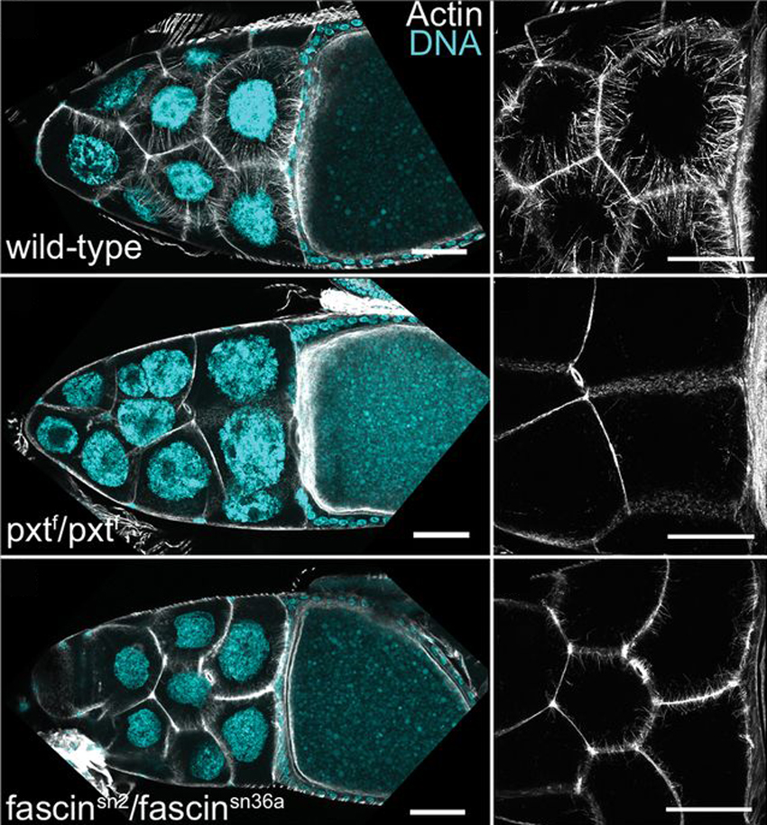
Prostaglandins (PGs) are lipid signaling molecules that regulate a wide range of processes, such as inflammation, pain, and hormone regulation. One target of PGs is the actin cytoskeleton, but how PGs affect actin filament polymerization and dynamics is not well understood. A recent paper uses the fruit fly egg chamber as a great model of how PGs regulate actin. In the fruit fly egg chamber, many nurse cells surround the oocyte (developing egg). These nurse cells are squeezed in order to dump all of their cytoplasmic contents through ring canals and into the growing egg. During this process, the nuclei of nurse cells are held in place by bundles of actin filaments to ensure that the nuclei don’t plug up the ring canals and block the transfer of the cytoplasm. PGs are an important part of this process, and they interact with an actin-bundling protein called Fascin, according to a recent paper by Groen and colleagues. Inhibition of either Fascin or Pxt, an enzyme required for PG production, results in the failure of actin bundle formation and nurse cell dumping. The loss of Fascin enhances the effects of Pxt reduction, and the overexpression of fascin suppresses the defects seen in flies with Pxt loss. Groen and colleagues show that PGs regulate Fascin, which modulates actin cytoskeleton rearrangements during nurse cell dumping. In the images above, control egg chambers (top) show parallel actin bundles (white) within nurse cells, which extend from the cell membranes to the nuclei (blue). pxt and fascin mutants (middle, bottom), however, contain little to no actin bundles in nurse cells.


ResearchBlogging.orgGroen, C., Spracklen, A., Fagan, T., & Tootle, T. (2012). Drosophila Fascin is a novel downstream target of prostaglandin signaling during actin remodeling Molecular Biology of the Cell, 23 (23), 4567-4578 DOI: 10.1091/mbc.E12-05-0417

October 19, 2012

The next time you see a fruit fly buzzing around your kitchen, take a beat before you smack it with a swatter and remind yourself of the amazing discoveries due to organisms like the (not so) lowly fruit fly. Maybe you’ll offer a thank-you glass of grape juice instead and show the intruder back outside. Today’s image is from a paper describing a fly model of a serious human disease, and serves as a great example of the power in a model organism.

Spinal muscular atrophy (SMA) is a heritable disease that results in infant mortality due motor neuron dysfunction and rapid degeneration of muscle. SMA is caused by the depletion of the SMN (survival motor neuron) protein. A recent paper describes the use of fruit flies in studying SMA, and shows that flies lacking SMN have reduced muscle size and defective motor neuron neurotransmission similar to SMA patients. Imlach and colleagues found that replenishing SMN levels in motor neurons and muscles did not reverse the defects of SMN depletion, yet increasing SMN levels in partner cells (proprioceptive neurons and interneurons) can reverse the defects. These results suggest that SMN depletion primarily affects the sensory-motor network, with secondary effects seen in the motor circuit. In addition, Imlach and colleagues found that increasing motor neural circuit excitability, either genetically or with drugs, could relieve SMN-depletion defects. Using this fly model of SMA, these results suggest that SMA patients may improve by enhancing motor neural network activity. The images above show the differences in muscle size in wild-type (left) and SMN-depleted (right) flies.


ResearchBlogging.orgImlach WL, Beck ES, Choi BJ, Lotti F, Pellizzoni L, & McCabe BD (2012). SMN Is Required for Sensory-Motor Circuit Function in Drosophila. Cell, 151 (2), 427-39 PMID: 23063130
Copyright ©2012 Elsevier Ltd. All rights reserved.


October 8, 2012

If you are a developmental biologist, there is a high probability that you study Wnt. The Wnt signaling pathway is employed throughout cell and developmental biology in processes ranging from spindle positioning to stem cell fate decisions. Today’s image is from a paper showing how Wnt can be secreted from cells.

Wnt signaling functions by relaying a signal from the cell’s surface to the nucleus, where gene expression is regulated. Active Wnt proteins are secreted out of a cell to induce tissue patterning during development (among many other things), and can travel over a distance of several cells. In trying to understand Wnt secretion, a recent paper describes results showing that active Wnt signals can be secreted from exosomes. Exosomes are vesicles that cells use to secrete various materials into the extracellular space. Gross and colleagues found that Wnt signals are secreted on exosomes in both developing fruit flies and human cells. Wnt signals are trafficked through various endosomal compartments and then to exosomes, with the help of the trafficking protein Ykt6 (an R-SNARE). In the top row of images above, Wnt (left column, red in merged) appears colocalized with an exosomal protein (CD63, green in merged) in developing fly wing discs. Wnt is also colocalized with a marker for multivesicular bodies (LAMP-1, green in merged), vesicles from which exosomes originate.  Insets show higher magnification views of the vesicles. 

ResearchBlogging.orgGross JC, Chaudhary V, Bartscherer K, & Boutros M (2012). Active Wnt proteins are secreted on exosomes. Nature cell biology, 14 (10), 1036-45 PMID: 22983114

Adapted by permission from Macmillan Publishers Ltd, copyright ©2012

September 24, 2012

If you are a mitotic spindle aficionado or superfan, then you understand how the mitotic spindle has a life of its own. Information on the spindle’s composition, dynamics, and list of duties can fill entire volumes…and there is still so much we are learning about the spindle. Today’s image is from a paper that adds another chapter in our metaphorical mitotic spindle book.

The mitotic spindle is composed of dynamic microtubules and countless proteins that ensure chromosomes are equally segregated into identical daughter cells at the end of cell division. Because much of the structure of the spindle comes from the cytoplasm, the contributions from the nucleus have remained unclear. A recent paper identifies the long-elusive presence of a nuclear matrix—a viscous matrix of proteins that supports the formation and function of the spindle. The nuclear matrix is composed of nuclear proteins that reassemble after nuclear envelope breakdown into a pole-to-pole structure. By looking at the cell divisions in fruit flies, Yao and colleagues found that the nuclear matrix is distinct from spindle microtubules, and is found closely around chromosomes following drug treatments that depolymerized microtubules. The time-lapse images above show mitosis in the syncytial Drosophila embryo. The nuclear matrix (green, Chromator) has a spindle-like morphology as the microtubule spindle (red, tubulin) itself forms. 


ResearchBlogging.orgYao C, Rath U, Maiato H, Sharp D, Girton J, Johansen KM, & Johansen J (2012). A nuclear-derived proteinaceous matrix embeds the microtubule spindle apparatus during mitosis. Molecular biology of the cell, 23 (18), 3532-41 PMID: 22855526

September 11, 2012

If I had to describe myself as a cell, I would want to say something about being a fired-up neuron, or a nurturing nurse cell, or a chameleon stem cell. Realistically, though, I’d be a muscle fiber—painfully organized and precise. When things aren’t so organized I’d just be a twitching little muscle cell until my closet was organized by sleeve length, my pantry in alphabetical order, my various to-do lists organized by priority. A recent paper describes how the nuclei and organelles within a muscle fiber are so regularly spaced (but sadly does not describe any suggestions for my own personal organization).

Striated muscle fibers have densely packed myofibrils, which are the structures that make muscle contractions happen. The muscle fiber is a large multinucleated cell, meaning that there are more than one nucleus. These nuclei, as well as other organelles, must be evenly distributed along the length of the fiber. KASH domain proteins play a role in positioning nuclei and other organelles in various cell types, and a recent paper found that two KASH proteins play this role in muscle cells in the fruit fly. Elhanany-Tamir and colleagues found that these two proteins, called Klar and MSP-300, cooperate to ensure the even spacing of nuclei within a muscle fiber. These two proteins link a nuclear ring of MSP-300 to astral microtubules. Without either of these KASH proteins, the nuclear spacing is irregular. MSP-300 also is important in the spacing of other organelles—mitochondria and ER—within the muscle fiber. In the images above, muscle fibers from wild-type (left),
msp-300 mutant, and klar mutant fruit fly larvae are stained for MSP-300 (red) and a nuclear marker (green). In the mutants, the MSP-300 nuclear ring (arrowheads) is dissociated from the nucleus.

ResearchBlogging.orgElhanany-Tamir H, Yu YV, Shnayder M, Jain A, Welte M, & Volk T (2012). Organelle positioning in muscles requires cooperation between two KASH proteins and microtubules. originally published in the Journal of Cell Biology, 198 (5), 833-46 PMID: 22927463

April 30, 2012

One might think that once you’ve won your first Nobel Prize, it’s time to kick back and watch the youngsters do the dirty work of solving all of biology’s riddles. No so. Apparently, Nobel-ers don’t like to kick back at all, but continue to push the limits of our knowledge. I think they’re all wearing superhero capes underneath their biologist uniforms (plaid shirts and old jeans?). Today’s image is from the lab of Eric Wieschaus, in a paper that provides a fascinating alternative mechanism to the prevailing model of epithelial folding.

During development, groups of cells are shaped into tissues and organs in a process called morphogenesis. One of the earliest morphogenesis events is gastrulation, during which the embryo is organized into the three germ layers that will each develop into different tissues and organs. During gastrulation in the fruit fly, the dorsal side of the embryo undergoes two epithelial folds, the anterior and posterior dorsal transverse folds, at precise locations on the embryo. The current model of epithelial folding involves actin-myosin contractions that trigger the initial cell shape changes. Wang and colleagues recently found an alternative mechanism that underlies epithelial folding during fruit fly gastrulation. Specifically, Wang and colleagues found that the adherens junctions that form between epithelial cells relocate to more basal locations in the cells that initiate epithelial folding. The movement of adherens junctions, and in turn dorsal fold initiation, depends on the activity of the polarity proteins Bazooka and Par-1. In the images above, the initiation of anterior (pink arrow) and posterior (blue arrow) folds are visualized by high resolution live imaging. The localization of E-cadherin (white), a key adherens junction protein, drops from the apical surface (top of the cells) to a more basal location in the cells undergoing shape changes.

ResearchBlogging.orgWang, Y., Khan, Z., Kaschube, M., & Wieschaus, E. (2012). Differential positioning of adherens junctions is associated with initiation of epithelial folding Nature, 484 (7394), 390-393 DOI: 10.1038/nature10938
Adapted by permission from Macmillan Publishers Ltd, copyright ©2012

December 12, 2011


If you have ever lived or worked with me (or are that poor guy who is married to me), then you know that I like things neat and organized. Anything less will send me into a sad tailspin that involves boxed wine and Cheetos. Thankfully, there are enough stunningly beautiful examples of precision, order, and patterning throughout biology to make me happy….like, really happy. Today’s image is from a paper that describes how the different cells in a fruit fly’s eye arrange into the honeycomb pattern seen above.

One of the big questions in developmental biology is how groups of different cell types arrange themselves to form a functional organ. A fantastic model to study this question is the compound fruit fly eye, made of hundreds of ommatidia. A group recently looked at how the several cell types in the developing fly eye are able to reorganize themselves into their honeycomb lattice. The very precise local movements of these cells, according to Johnson and colleagues, require regulation by a protein called Arf6 GTPase in order to connect cell surface signaling with the cytoskeletal rearrangements required for cell motility. The adaptor protein, Cindr, is able to bind to and sequester Arf6 activators called ArfGAPs, which in turn prevents local Arf6 activity. Images above show the precise honeycomb organization in a normal fruit fly pupal eye. In the developing eye (shown chronologically from left to right), the cone cells (orange in cartoon) of each ommatidia are surrounded by a hexagon lattice of cells.

ResearchBlogging.orgJohnson, R., Sedgwick, A., D'Souza-Schorey, C., & Cagan, R. (2011). Role for a Cindr-Arf6 axis in patterning emerging epithelia Molecular Biology of the Cell, 22 (23), 4513-4526 DOI: 10.1091/mbc.E11-04-0305

November 28, 2011

Asymmetric cell division results in two unequal daughter cells and without it, stem cells would not hold the attention of biologists, sick patients, politicians, and those loud nut-jobs who have more passion than knowledge. Check out today’s image, from a paper identifying a new protein complex important in asymmetric division.

Asymmetric division occurs throughout development, and is when a cell divides to result in two daughter cells that are unequal in size and/or cell fate. Part of this process is the establishment of the axis along which the cell will divide. Once this polarity is established, the mitotic spindle can line up accordingly. Fruit fly neural stem cells, called neuroblasts, divide asymmetrically to result in the birth of another neuroblast and a smaller ganglion mother cell, which eventually gives rise to neurons. A recent paper identifies a new role for a signaling complex in establishing neuroblast polarity. In this paper, Carmena and colleagues found that the Rap1–Rgl–Ral complex of proteins regulates polarity establishment in fruit fly neuroblasts, and works alongside other well-studied polarity proteins such as aPKC, Par6, and Pins. As seen in the images above, Rap1 (bottom row, green in merged; arrows) is enriched on only one side (apical side) of neuroblasts throughout division (DNA is in red).

ResearchBlogging.orgCarmena, A., Makarova, A., & Speicher, S. (2011). The Rap1-Rgl-Ral signaling network regulates neuroblast cortical polarity and spindle orientation originally published in The Journal of Cell Biology, 195 (4), 553-562 DOI: 10.1083/jcb.201108112

November 10, 2011

Mistakes happen. If you are as special as DNA, then you have someone to point out those mistakes and fix them for you. That sounds like your PI editing your manuscript, doesn’t it? Today’s stunning image is from a paper describing DNA repair in meiotic divisions.

When double-strand breaks happen to DNA, the cell has an efficient pathway that recognizes and repairs the breaks to both DNA strands. Two kinase proteins, ATM and ATR, play pivotal roles by phosphorylating numerous key proteins involved in this process. The roles of these two proteins are well-studied in mitosis, but their function in meiosis is not clear. Meiosis is the reductive cell division that results in gametes (ooctyes or sperm). A recent paper describes the role of ATM and ATR in the meiotic cell divisions of fruit fly ovaries. Both ATM and ATR phosphorylate a protein called histone H2AV at the site of DNA breaks, providing a handy and fluorescently-labeled output of ATM and ATR activity. Joyce and colleagues found that ATR plays a role in regulating the cell cycle checkpoint machinery that halts cell cycle progression in the presence of DNA breaks, while ATM is required for the DNA repair of meiotic double strand breaks. As seen in the images above, H2AV levels (red, phosphorylated and unphosphorylated) decreased in the developing oocyte (green) until it was almost undetectable by stages 4 and 5 of oogenesis (see bottom row for higher magnification). These results suggest that either ATM/ATR does not respond to DNA damage at these stages or that repair occurs before these stages (before the first meiotic division).

ResearchBlogging.orgJoyce, E., Pedersen, M., Tiong, S., White-Brown, S., Paul, A., Campbell, S., & McKim, K. (2011). Drosophila ATM and ATR have distinct activities in the regulation of meiotic DNA damage and repair originally published in The Journal of Cell Biology, 195 (3), 359-367 DOI: 10.1083/jcb.201104121

November 7, 2011

Actin is that super achieving big man-or-woman on campus who seems to be involved in and excelling at everything. Even if you roll your eyes at actin’s achievements and wonder what actin ISN’T involved in, you’re secretly impressed and jealous. Today’s image is from a paper on axon guidance and the role of the actin regulators WAVE and WASP.

Like many developmental processes, the guidance and mobility of developing axons depends on a dynamic actin cytoskeleton. Because most neuronal networks are quite complex, the use of the fruit fly’s eye provides biologists with a genetically-tractable model for understanding axon growth and guidance. The fly’s eye is a compound eye—there are 750 individual eyes called ommatidia, each comprised of 8 light-sensitive photoreceptor neurons (R-cells). A recent study tests the roles of two actin nucleating regulators called WASP and WAVE in R-cell axon targeting. In this study, Stephan and colleagues found that a protein called Abi regulates WAVE by ensuring its membrane localization, where WAVE activates the Arp2/3 actin nucleating complex. While WAVE is required for R-cell axon guidance, WASP is not. The images above show R-cell axons (green) in flies with different genetic backgrounds. Wild-type flies (left) and
wasp mutants (right) have regular patterns and spacing of axons, while wave mutants (middle) have bundled or clumped axons with irregular spacing.

ResearchBlogging.orgStephan, R., Gohl, C., Fleige, A., Klambt, C., & Bogdan, S. (2011). Membrane-targeted WAVE mediates photoreceptor axon targeting in the absence of the WAVE complex in Drosophila Molecular Biology of the Cell, 22 (21), 4079-4092 DOI: 10.1091/mbc.E11-02-0121