June 25, 2013

Maybe you’ve been cruising in your minivan blasting “The Wheels on the Bus” too loudly for your preschooler, or maybe you’re preparing to hear the witching hour’s cries of a newborn due soon (this blog is way too autobiographical).  Either way, our ears endure a lot of stress from loud sounds throughout our lives.  The images above are from a paper describing a mechanism for handling this stress.

Our inner ear hair cells detect sound when stereocilia projections are deflected by sound vibrations.  These actin-based projections are connected to each other by tiny extracellular tip links, which are tugged when the stereocilia bend.  The tugging of these tip links is believed to drive the opening of transduction channels that result in the electrical signals relayed to the auditory nerve.  Although mammalian hair cells do not regenerate over the course of a lifetime, the tip links do.  A recent paper investigates the mechanism behind tip link regeneration.  Indzhykulian and colleagues developed an electron microscopy technique to visualize the tip links on hair cell stereocilia and immuno-gold labeled proteins of tip links, and found a two-step mechanism for tip link regeneration.  This mechanism begins with shorter tip links composed of just protocadherin 15, and follows with mature tip links containing protocadherin 15 and cadherin 23.  In the images above, stereocilia tip links were regenerated after nearly complete tip disruption by the chemical BAPTA.  Insets show higher magnified views of the tip links, which appear regenerated by 24-48 hours, compared with control tip links (top) and BAPTA-treated cells (second row).

ResearchBlogging.orgIndzhykulian AA, Stepanyan R, Nelina A, Spinelli KJ, Ahmed ZM, Belyantseva IA, Friedman TB, Barr-Gillespie PG, & Frolenkov GI (2013). Molecular remodeling of tip links underlies mechanosensory regeneration in auditory hair cells. PLoS biology, 11 (6) PMID: 23776407

June 21, 2013

Good things come in small packages.  Maybe I’m referring to the burst of antioxidants jammed into tiny blueberries.  Maybe I’m referring to my tiny three year-old who yells, “Come oooon, THAT was funny!” when I don’t laugh loudly enough at her jokes.  Or maybe I’m referring to C. elegans.  These worms are tiny, but pack a serious punch of significant biology that helps us learn about important cellular processes.  Today’s image is from a paper that serves as an excellent example of this.

During cell invasion, a cell is able to breach and cross over the basement membrane that underlies a sheet of epithelial cells.  Cell invasion occurs throughout development and in the spread of cancer, yet biologists studying cell invasion have been challenged by the difficulty of visualizing the event.  A recent paper describes the development of live-cell imaging methods for studying cell invasion, using the worm’s anchor cell.  The anchor cell in the developing worm’s uterus breaches the basement membrane in order to link uterine and vulval tissues, and its transmigration is precisely timed.  Hagedorn and colleagues followed the interactions between the invading anchor cell and the basement membrane, and found very dynamic actin-based invadopodia that first breach the basement membrane.  These protrusions then stabilize to expand the breach and cross into the vulval tissue.  Anchor cell invasion depends on the netrin receptor UNC-40 (DCC) at the interface between the anchor cell and basement membrane.  In the time-lapse images above, the invading protrusion (cyan in top, grayscale in bottom) can be seen breaching the basement membrane (purple) and invading the vulval tissue underneath.

ResearchBlogging.orgHagedorn, E., Ziel, J., Morrissey, M., Linden, L., Wang, Z., Chi, Q., Johnson, S., & Sherwood, D. (2013). The netrin receptor DCC focuses invadopodia-driven basement membrane transmigration in vivo originally published in the Journal of Cell Biology, 201 (6), 903-913 DOI: 10.1083/jcb.201301091

June 18, 2013

The study of how cells move in development is not just about development.  Understanding cell migration can also help researchers understand how tumors spread and invade other tissues.  So, the next time you see someone roll their eyes at your fruit fly egg chambers (or worm vulva, or culture dishes), take pity at their ignorance and explain to them how they should thank you instead.

The movement of cells during development drives the shape changes and organization of an embryo.  In the fruit fly ovary, a small cluster of border cells migrates across a region of the egg chamber in order to reach the oocyte.  This collective migration of these border cells depends on polarization of the actin cytoskeleton.  A recent paper describes the role of the Hippo signaling pathway in driving the polarization of actin to the outer rim of the migrating border cell cluster.  Lucas and colleagues found that upstream Hippo pathway components localize to the contacts between border cells within the cluster in order to link polarity signaling with actin cytoskeleton organization.  In the images above, the actin cytoskeleton (red) can be seen at the outer rim of the migrating cluster of border cells (arrows) as it moves across the egg chamber towards the oocyte (top to bottom, chronologically).  Higher magnification views of the cluster are on the right.

 ResearchBlogging.orgLucas, E., Khanal, I., Gaspar, P., Fletcher, G., Polesello, C., Tapon, N., & Thompson, B. (2013). The Hippo pathway polarizes the actin cytoskeleton during collective migration of Drosophila border cells originally published in the Journal of Cell Biology, 201 (6), 875-885 DOI: 10.1083/jcb.201210073

June 11, 2013

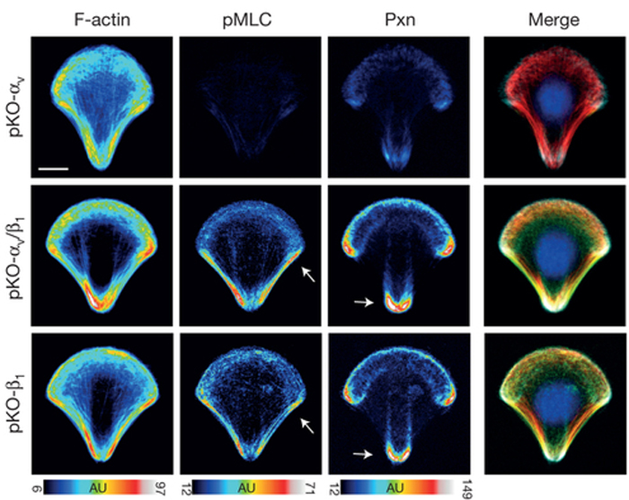
 I’m willing to bet that most scientists were pretty destructive as kids.  Not intentionally destructive, though…I bet we all liked taking things apart to see what each part of a toy did.  Maybe your Teddy Ruxpin eventually sounded like a demonic doll after your experiments, or you finally pulled apart your Etch-A-Sketch to uncover the magic.  Either way, it was early training for what scientists do every day to understand cells better.  Today’s image is from a paper that takes this route to understand cell adhesion better.

Integrins are transmembrane proteins that attach a cell to another cell or its surrounding extracellular matrix, so their role in regulating cell adhesion in normal cell function, development, and disease is of serious interest to biologists.  There are several types of integrins, frequently expressed together at the same time.  A recent paper describes a system to investigate how specific integrins function in cell adhesion, and found notable differences.  Schiller and colleagues expressed either β1- or αv-class integrins in fibroblasts lacking all other integrins, and found that β1-class integrins are important in small peripheral adhesions, while αv-class integrins are important for large focal adhesions.  In the images above, activity of the actin-based motor myosin and presence of the focal adhesion protein paxillin are shown in cells plated on micropatterned shapes.  Cells with both β1- and αv-class integrins (middle row) have the highest myosin activity (pMLC) and paxillin signal, while cells with either β1- or αv-integrin alone have lower myosin activity and paxillin signal.  

ResearchBlogging.orgSchiller, H., Hermann, M., Polleux, J., Vignaud, T., Zanivan, S., Friedel, C., Sun, Z., Raducanu, A., Gottschalk, K., Théry, M., Mann, M., & Fässler, R. (2013). β1- and αv-class integrins cooperate to regulate myosin II during rigidity sensing of fibronectin-based microenvironments Nature Cell Biology, 15 (6), 625-636 DOI: 10.1038/ncb2747
Adapted by permission from Macmillan Publishers Ltd, copyright ©2013 

June 4, 2013

HighMag is back from an early summer vacation at the beach, and ready to get our microscopic groove on.  Happy Summer, everyone!


Our world is the same size it’s always been, but so many advances in technology have made the world seem a lot smaller.  We can call our brother across the country and video chat with our sister on the other side of the world, all while searching the internet for the lyrics to Snow’s “Informer” (side note…knowing the lyrics won’t help you understand the song AT ALL).  Similarly, a neuron’s axons make the nervous system seem a lot smaller too, with their amazing ability to grow very long.  Today’s image is from a paper showing how microtubule sliding is involved.

Neurons can transmit signals to far away neurons, thanks to the ability of their axons to grow insanely long.  Both actin filaments and microtubules participate in axon growth, with microtubules pushing the growing axon out and actin filaments working directly at the tip of the growing axon, called the growth cone.  A recent paper shows the role of microtubule sliding in the initial growth of neurites in fruit fly neurons.  Lu and colleagues found that the microtubule motor kinesin-1 drives the sliding of microtubules past one other in order to push out the neuron’s growing projection.  This mechanism does not require actin filaments, and is suppressed during maturation of neurons.  In the images above, microtubules (green) can be seen pushing against the tip of a growing neurite in a young neuron (membrane of neuron in red and bottom right series).  The whole neuron is on the left, and the neurite in the boxed region is shown in the time-lapse images on the right.

ResearchBlogging.orgLu, W., Fox, P., Lakonishok, M., Davidson, M., & Gelfand, V. (2013). Initial Neurite Outgrowth in Drosophila Neurons Is Driven by Kinesin-Powered Microtubule Sliding Current Biology, 23 (11), 1018-1023 DOI: 10.1016/j.cub.2013.04.050 
Copyright ©2013 Elsevier Ltd. All rights reserved.