Today’s image is from a paper describing a new microscopy method that allows the best of both worlds – fluorescent AND electron microscopy. The images in this paper are fantastic, but I especially can’t wait to see what images are down the road using this new method.
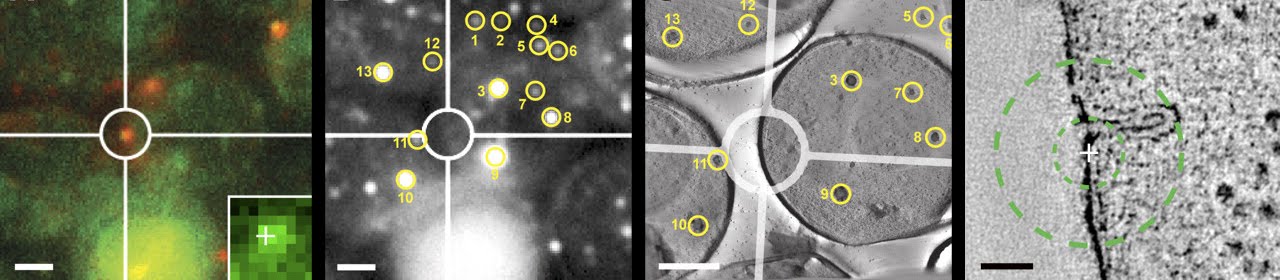
The strength of electron microscopy lies in its ability to reveal amazing detail about cellular structure, yet transient and dynamic events are difficult to see using this method. Fluorescence microscopy, however, is invaluable in the visualization of dynamic events, even if it cannot provide structural detail. A recent paper has combined the best of both techniques in a correlative fluorescence and electron microscopy method that provides high precision and sensitivity. In the image above, a yeast endocytic protein was imaged using fluorescence microscopy (red mark inside the circle, left-most image). Fiducial markers (numbered circles) in both fluorescence (middle left) and electron micrographs (middle right) help align the images, in short. Finally, a high magnification electron micrograph (right-most image) shows the structural detail of a membrane invagination where the endocytic protein was found in the fluorescence image (marked with a cross).
No comments:
Post a Comment