Showing posts with label actin. Show all posts

March 26, 2015

The actin cytoskeleton at the leading edge of a crawling cell has been a source of both scientific fascination and stunning images for biologists. Today’s image is from a paper that sheds light on how that complex structure is generated. 

The outermost region of a crawling cell’s cortex is called the lamellipodium, and it depends on a complex actin cytoskeleton for its structure and rapid dynamics. The generation of branched actin filaments at lamellipodia requires the activity of the actin-nucleating Arp2/3 protein complex. In a recent study, Henson and colleagues use the very flat and Arp2/3-rich lamellipodia of sea urchin coelomocytes to visualize the actin cytoskeleton. As seen in the images above, treatment of coelomocytes with a drug that inhibits the Arp2/3 complex resulted in drastic changes to the actin cytoskeleton. Specifically, the densely-packed branched filaments of lamellipodia (left, control) were replaced with actin filament arcs that ran diagonal or parallel to the cell’s edge (30 and 60 seconds after drug treatment, middle and right images). These transverse actin arcs may act as mother/scaffold filaments from which more actin filaments are nucleated during organization of the cytoskeleton in lamellipodia.

Henson, J., Yeterian, M., Weeks, R., Medrano, A., Brown, B., Geist, H., Pais, M., Oldenbourg, R., & Shuster, C. (2015). Arp2/3 complex inhibition radically alters lamellipodial actin architecture, suspended cell shape, and the cell spreading process Molecular Biology of the Cell, 26 (5), 887-900 DOI: 10.1091/mbc.E14-07-1244

January 8, 2015

If you are lucky in life, there is at least one person who will always be there for you—a parent, your spouse, maybe even your pooch. As we understand more and more of what goes on inside a cell, it has become clear that actin is always there for the cell’s many organelles. Actin is so supportive and encouraging, and without it our cells would just be puddles of fats and proteins. Today’s images are from a paper describing the role of actin in mitochondrial fission. 

Mitochondria are dynamic organelles that divide by fission. Although a role for the actin cytoskeleton in mitochondrial fission has been suggested, the exact mechanism is unclear. Recent work by Li and colleagues shows a transient association of F-actin (filamentous actin) to mitochondria at the start of fission. Downregulation of the actin regulators cortactin, cofilin, and Arp2/3 caused elongation of mitochondria. Li and colleagues tested the role of Drp1, which is a key player in mitochondrial division, on F-actin assembly during fission. Drp1 inhibition prolonged the localization of F-actin and several actin regulators at mitochondria during fission. In the left group of images above, F-actin (green) and mitochondria (red) are visible in a control mammalian cell (bottom row is at higher magnification). The group of images on the right shows mammalian cells after chemical induction of mitochondrial fission: 2 minutes after drug treatment, many F-actin-rich mitochondria are visible.

Li, S., Xu, S., Roelofs, B., Boyman, L., Lederer, W., Sesaki, H., & Karbowski, M. (2014). Transient assembly of F-actin on the outer mitochondrial membrane contributes to mitochondrial fission The Journal of Cell Biology, 208 (1), 109-123 DOI: 10.1083/jcb.201404050

September 5, 2014

It is so nice to have a friend who truly complements you…someone similar to you, but different enough to pick up the slack of your own shortcomings. Today’s image is from a paper about the Laverne and Shirley partnership of Ena/VASP and mDia2. 

Crawling cells extend finger-like filopodia to probe the environment for cues and to establish adhesion of the cell to the substrate. Filopodia are composed of parallel bundles of actin that are quickly dynamic. Countless actin regulators affect filopodia formation, some of which have seemingly similar functions. The Enabled (Ena)/VASP and Diaphanous 2 (mDia2) proteins are both actin polymerases, but as a recent paper by Barzik and colleagues describes, they support filopodia formation in distinct, non-redundant ways. By using mouse embryonic fibroblasts lacking both Ena/VASP and mDia2, Barzik and colleagues found that filopodia formed using either Ena/VASP or mDia2 alone differed in number, actin filament organization, lifetime, and other parameters. Filopodia generated using mDia2 alone were not able to initiate integrin-dependent adhesion and lamellipodial protrusions. The image above shows a cell with both mDia2 (red) and Ena/VASP (green), with the two proteins colocalizing on a subset of filopodia (arrows).

Barzik, M., McClain, L., Gupton, S., & Gertler, F. (2014). Ena/VASP regulates mDia2-initiated filopodial length, dynamics, and function Molecular Biology of the Cell, 25 (17), 2604-2619 DOI: 10.1091/mbc.E14-02-0712

April 30, 2014

Have you ever driven in the wrong direction on a one-way street. It feels as wrong as a hamburger smoothie and you feel overwhelmed with panic. It’s important to go the right direction on one-way streets, and a neuron understands this. Neurons are polarized so that signals can come and go in the right direction. Today’s stunning image is from a paper describing the cytoskeletal architecture within a region of a neuron that’s important for polarity. 

The axon initial segment (AIS) is the part of an axon closest to the neuron’s cell body, and is the site of action potential initiation. The AIS is crucial for the neuron’s polarity, which facilitates the direction of incoming signals (coming in from dendrites) and outgoing information (out along the axon to the synapse). A recent study from Jones and colleagues investigated how the AIS maintains neuronal polarity. Jones and colleagues used platinum replica electron microscopy (PREM) to image the cytoskeleton in hippocampal neurons, and found that it begins with a bundle of microtubules. A dense fibrillar–globular coat covers this microtubule bundle and contains many proteins as well as actin filaments. Actin filaments are found in two sparse populations—either stable, short filaments or dynamic, long filaments. Jones and colleagues propose that the dynamic actin filaments play a role in the AIS coat, while the stable filaments may play a structural role in the AIS diffusion barrier. This diffusion barrier prevents the mixing of plasma membrane components from dendrites and axons, an important factor in maintaining polarity. The image above shows microtubules within the AIS, with thin fibrils (arrows) and a fibrillar coat over the microtubules (arrowheads) visible.

Jones, S., Korobova, F., & Svitkina, T. (2014). Axon initial segment cytoskeleton comprises a multiprotein submembranous coat containing sparse actin filaments originally published in the Journal of Cell Biology, 205 (1), 67-81 DOI: 10.1083/jcb.201401045

April 24, 2014

I’m still waiting for my miniaturizing spaceship* so I can dive into a cell with my dog and ride in the lamella of a crawling cell. Until then, I will gladly enjoy images like today’s, from the Lippincott-Schwartz lab.

A cell’s shape can change for many reasons, including crawling, tissue regeneration, and cancer progression. Cell shape is dynamic, relying on temporal and spatial coordination of several processes. The three-dimensional nature of cell shape, however, presents a challenge for microscopists. By using structured illumination microscopy (SIM), Burnette and colleagues created images of the three-dimensional organization of actin filaments. From these 3D superresolution analyses in crawling cells, Burnette and colleagues found a contractile network of actin filaments at the top of a crawling cell, organized like muscle sarcomeres. Their model of balanced contraction and adhesion helps to guide further investigations of cell shape changes in healthy and diseased tissue. The image above shows actin filaments at color-coded heights within a crawling cell. The closed arrow points to actin at the top of the cell.

*My birthday is next month, if anyone is wondering about gift ideas.  

Burnette, D., Shao, L., Ott, C., Pasapera, A., Fischer, R., Baird, M., Der Loughian, C., Delanoe-Ayari, H., Paszek, M., Davidson, M., Betzig, E., & Lippincott-Schwartz, J. (2014). A contractile and counterbalancing adhesion system controls the 3D shape of crawling cells originally published in the Journal of Cell Biology, 205 (1), 83-96 DOI: 10.1083/jcb.201311104

April 17, 2014

The endoplasmic reticulum and humans have quite a bit in common. Both are dynamic and constantly changing, but both also need something to ground and stabilize them. Maybe I’m reading too much into the beauty of the ER, but the image today is from a paper that only fuels my fascination.

The endoplasmic reticulum (ER) is a large, complex membrane-bound organelle that spreads throughout the cell and hosts the synthesis, folding, and sorting of membrane and secretory proteins. This network is dynamic and constantly rearranging, with diverse structural and functional domains. A recent paper describes the investigation into the role of the actin cytoskeleton in regulating ER sheet persistence and maintenance, which is important as the stationary domain of the ER. Joensuu and colleagues identified a subset of actin filaments associated with the ER, specifically to polygons defined by ER tubules and sheets. Actin depolymerization caused ER sheet fluctuation and resulted in a defective ER network. The actin motor myosin 1c also localizes to these actin filament arrays. In the images above, actin filament arrays (magenta, arrows) localize to the polygons (asterisks) associated with the ER (green).

Joensuu, M., Belevich, I., Ramo, O., Nevzorov, I., Vihinen, H., Puhka, M., Witkos, T., Lowe, M., Vartiainen, M., & Jokitalo, E. (2014). ER sheet persistence is coupled to myosin 1c-regulated dynamic actin filament arrays Molecular Biology of the Cell, 25 (7), 1111-1126 DOI: 10.1091/mbc.E13-12-0712

April 10, 2014

When you host a party at your home, do you hire a caterer to bring in food or do you cook the food right there in your kitchen? One of these options leaves a lot more wiggle room for last-minute changes—a few extra guests, a gluten allergy, a pregnant lady with a disgust for wobbly desserts. A cell recognizes this distinction too. When making certain proteins, a cell will synthesize proteins where and when they’re needed. Today’s image is from Natasha Gutierrez, who recently published a study describing the role of β-actin mRNA and monomer synthesis in adherens junction assembly.

The actin cytoskeleton is made of actin filaments and countless actin-regulating proteins that guide the ever-changing dynamics of the cytoskeleton. Actin filament polymerization is regulated by localized synthesis of β-actin monomers from β-actin mRNA. A recent paper by Gutierrez and colleagues shows that the assembly of adherens junctions (AJs), epithelial cell-cell adhesion structures, requires localized β-actin monomer synthesis, the β-actin 3’ UTR and β-actin mRNA zipcode sequence at cell-cell contact sites. Additionally, active RhoA, which targets zipcode-mediated β-actin mRNA localization, is localized to cell-cell contact sites. In the unpublished images above, mammalian cells were treated with increasing levels (top to bottom) of a Rho inhibitor. The ability to form AJs, seen as the colocalization of actin filaments (left, green in merged) and E-cadherin (middle, red in merged) at cell-cell contact sites, decreased with increasing levels of the Rho inhibitor.

ResearchBlogging.orgGutierrez, N., Eromobor, I., Petrie, R., Vedula, P., Cruz, L., & Rodriguez, A. (2014). The B-actin mRNA zipcode regulates epithelial adherens junction assembly but not maintenance RNA DOI: 10.1261/rna.043208.113

February 5, 2014

The next time you try swatting away that little fruit fly from a neighboring lab while you enjoy your midday coffee break, take a beat and appreciate how stinkin’ purrrty those flies are. Today’s image features the developing egg of the fruit fly, and accompanies a paper describing the important role for prostaglandins in the (very photogenic) process.

Prostaglandins (PGs) are small lipids that act as signaling molecules in various physiological processes such as pain, inflammation, and platelet aggregation. On the cellular level, PGs regulate the organization of the actin cytoskeleton. A recent paper in Molecular Biology of the Cell from the Tootle lab sheds light on the role of PGs in development, and shows that PGs temporally regulate actin cytoskeleton organization during Drosophila oogenesis. Specifically, PGs function at stages 9 and 10 of oogenesis to inhibit, then promote, actin remodeling via the actin elongation factor Ena. Loss of PG signaling at stage 9 triggers early actin filament formation and bundling, while loss of PG signaling at stage 10 triggers a reduction of, or complete loss of, parallel actin filament bundling. As seen in the images above, actin filament organization (white) is abnormal in two different PG mutant follicles (middle, bottom), compared to a wild-type follicle (top).

Andrew J. Spracklen, Daniel J. Kelpsch, Xiang Chen, Cassandra N. Spracklen, & Tina L. Tootle (2014). Prostaglandins temporally regulate cytoplasmic actin bundle formation during Drosophila oogenesis Molecular Biology of the Cell, 25 (3) DOI: 10.1091/mbc.E13-07-0366

January 29, 2014

The term “pathogen propulsion” sounds like an awesome technique for defeating the evil squid overlords. In fact, a lot of concepts involving propulsive actin comets sound awesomely science fictional, but thankfully they are not. Today’s image is from a paper describing how several viruses use actin comet tails to propel themselves to other cells.

Several pathogens such as baculovirus, Listeria, and Shigella hijack their host cell’s own actin cytoskeleton in order to propel themselves into other cells and spread infection. Behind each pathogen is a comet made of actin filaments and associated actin regulators, but the mechanism of propulsion and the structure of the actin comet have been debated. A recent paper in PLoS Biology by Mueller and colleagues describes the use of electron tomography to show a fishbone-like array of actin filaments behind baculovirus, the smallest pathogen known to use propulsive actin comets. These comets use an average of four actin filaments at any one time to propel the virus. Using these results, the researchers ran computer simulations that support a model of propulsion in which actin filaments are continuously tethered to the pathogen. The image above shows a negatively-stained actin comet tail behind a baculovirus particle (BV), and the 3D projection of the image shows branch points of the actin tail as red dots. Insets in top image show details of the branch points, and grey tube is a microtubule.

BONUS! Below is a movie of baculovirus propelling itself around a cell. Virus particles are red, and actin comet tails can be seen in green behind the particles.

Mueller J, Pfanzelter J, Winkler C, Narita A, Le Clainche C, et al. (2014) Electron Tomography and Simulation of Baculovirus Actin Comet Tails Support a Tethered Filament Model of Pathogen Propulsion. PLoS Biol 12(1): e1001765. doi:10.1371/journal.pbio.1001765.

July 17, 2013

We don’t need to reinvent the wheel (even if someone tried to in 2001...click here).  We use the wheel for so many things ranging from transport to energy.  Cells have proven clever at co-opting machinery for multiple processes, as the paper from today’s image describes.  This recent paper shows the use of specific machinery in both cytokinesis and neuronal migration.

When neurons migrate, there is a leading process in the front of the cell body and a trailing process.  The leading process contains actin filaments that enable the cell body of the neuron to move forward.  A recent paper describes how the microtubule-based motor kinesin-6 plays an important role in neuronal migration.  Kinesin-6 is best known for its role in cytokinesis, the physical division of a cell after mitosis.  Falnikar and colleagues found that kinesin-6 concentrates in the same region as actin filaments in the leading process of a migrating neuron.  Without kinesin-6, neurons lose their bipolar leading-trailing process morphology, concentrate actin filaments in more than one process, and either remain stationary or continually change the direction of migration.  In addition, Falnikar and colleagues found that kinesin-6 signals through the GTPase activating protein MgcRacGAP to regulate the actin cytoskeleton, as it does during cytokinesis.  In the images above, control neurons (top time-lapse series) moved in a single direction, while neurons depleted of kinesin-6 bottom) frequently changed directions.

BONUS!!  Check out a movie of a wandering, migrating kinesin-6-depleted neuron below.

ResearchBlogging.orgAditi Falnikar, Shubha Tole, Mei Liu, Judy S. Liu, & Peter W. Baas (2013). Polarity in Migrating Neurons Is Related to a Mechanism Analogous to Cytokinesis Current Biology, 23 (13), 1215-1220 DOI: 10.1016/j.cub.2013.05.027 Copyright ©2013 Elsevier Ltd. All rights reserved.

July 10, 2013

The grace of a migrating cell is as deceiving as a pair of Spanx on an English Bulldog… there is a lot going underneath.  Today’s image is from a paper showing the importance of the protein vinculin at the leading edge of a migrating cell.

Cell migration is driven by actin filament polymerization that pushes the leading edge of the cell forward, as well as F-actin retrograde flow.  Focal adhesions (FAs) adhere the crawling cell to the underlying extracellular matrix (ECM), and are assembled and disassembled near the leading edge of the cell.  Proteins of these FAs are believed to make up a “molecular clutch” that engages the retrograde F-actin flow, and a recent paper identifies the protein vinculin, an actin-binding protein, as a molecular clutch.  Thievessen and colleagues investigated the effects of vinculin gene disruption in migrating fibroblasts, and found that vinculin is important in regulating F-actin organization and FA dynamics.  Specifically, vinculin generates the ECM traction forces necessary for migration, and promotes FA formation and turnover.  In the images above, a normal fibroblast (top) and a fibroblast lacking vinculin (bottom) show F-actin (green) and the lamellipodial protein cortactin (purple).  Normal crawling fibroblasts have a sharply defined band of cortactin colocalized with F-actin at the leading edge, while vinculin mutants have a wider, less defined region of cortactin at the leading edge, suggesting the importance of vinculin in leading edge organization.

BONUS!  Check out some cool movies from this paper here.

ResearchBlogging.orgThievessen I, Thompson PM, Berlemont S, Plevock KM, Plotnikov SV, Zemljic-Harpf A, Ross RS, Davidson MW, Danuser G, Campbell SL, & Waterman CM (2013). Vinculin-actin interaction couples actin retrograde flow to focal adhesions, but is dispensable for focal adhesion growth. originally published in the Journal of Cell Biology, 202 (1), 163-77 PMID: 23836933

June 21, 2013

Good things come in small packages.  Maybe I’m referring to the burst of antioxidants jammed into tiny blueberries.  Maybe I’m referring to my tiny three year-old who yells, “Come oooon, THAT was funny!” when I don’t laugh loudly enough at her jokes.  Or maybe I’m referring to C. elegans.  These worms are tiny, but pack a serious punch of significant biology that helps us learn about important cellular processes.  Today’s image is from a paper that serves as an excellent example of this.

During cell invasion, a cell is able to breach and cross over the basement membrane that underlies a sheet of epithelial cells.  Cell invasion occurs throughout development and in the spread of cancer, yet biologists studying cell invasion have been challenged by the difficulty of visualizing the event.  A recent paper describes the development of live-cell imaging methods for studying cell invasion, using the worm’s anchor cell.  The anchor cell in the developing worm’s uterus breaches the basement membrane in order to link uterine and vulval tissues, and its transmigration is precisely timed.  Hagedorn and colleagues followed the interactions between the invading anchor cell and the basement membrane, and found very dynamic actin-based invadopodia that first breach the basement membrane.  These protrusions then stabilize to expand the breach and cross into the vulval tissue.  Anchor cell invasion depends on the netrin receptor UNC-40 (DCC) at the interface between the anchor cell and basement membrane.  In the time-lapse images above, the invading protrusion (cyan in top, grayscale in bottom) can be seen breaching the basement membrane (purple) and invading the vulval tissue underneath.

ResearchBlogging.orgHagedorn, E., Ziel, J., Morrissey, M., Linden, L., Wang, Z., Chi, Q., Johnson, S., & Sherwood, D. (2013). The netrin receptor DCC focuses invadopodia-driven basement membrane transmigration in vivo originally published in the Journal of Cell Biology, 201 (6), 903-913 DOI: 10.1083/jcb.201301091

May 13, 2013

Molecular motors are some of the raddest things in a cell.  They can walk along cytoskeletal elements such as microtubules and actin filaments, and the list of cellular events that they participate in is a long, long list.  Today’s image is from a paper showing a beautiful pattern of nonmuscle myosin II in epithelial cells.

Epithelial cells assemble junctions to adhere to one another, and the actin motor nonmuscle myosin II (NMII) is a major component of these epithelial apical junctions.  NMII helps the epithelial sheet respond to morphogenesis and changes in tissue homeostasis, and a recent paper describes how the network of NMII motors does so.  Ebrahim and colleagues have found that NMII in the apical junctional complex of epithelial cells assembles into precise muscle-like sarcomere units that form a belt around each cell.  The sarcomeres of neighboring cells are aligned, in turn assembling into a contractile network that can result in changes in cell shape.  In the images above, NMII (green) is seen in repeated sarcomere units around each cell (actin is in red).  NMII puncta are paired together in neighboring cells.  Arrows (middle) point to the junctions between three cells, seen at higher magnification on the right.

ResearchBlogging.orgEbrahim, S., Fujita, T., Millis, B., Kozin, E., Ma, X., Kawamoto, S., Baird, M., Davidson, M., Yonemura, S., Hisa, Y., Conti, M., Adelstein, R., Sakaguchi, H., & Kachar, B. (2013). NMII Forms a Contractile Transcellular Sarcomeric Network to Regulate Apical Cell Junctions and Tissue Geometry Current Biology, 23 (8), 731-736 DOI: 10.1016/j.cub.2013.03.039 
Copyright ©2013 Elsevier Ltd. All rights reserved.

May 9, 2013

When we think of wounds, we don’t typically think of them as part of normal, healthy function.  Micro-wounds, however, form when white blood cells have to cross the barrier in our blood vessels to get to an injury or infection.  These micro-wounds happen all the time, and our cells heal these wounds efficiently and elegantly.

One of the most important barriers in our body is that created by the vascular endothelium.  Vascular endothelial cells line all of our blood vessels—from the largest vessels to the smallest capillaries—and function in fluid filtration, hormone trafficking, and recruitment and trafficking of blood and stem cells.  The movement of cells, for example white blood cells, across the vascular endothelium and out of circulation creates “micro-wounds” that can compromise the integrity of the tissue.  A recent paper describes how these micro-wounds are healed, based on a model in which the vascular endothelium senses a loss of tension upon micro-wounding and triggers its own repair.  Martinelli and colleagues tracked micro-wounds that were created by either transmigrating white blood cells or by mechanical disruption by a probe, and found that ventral lamellipodia are generated by endothelial cells to close the micro-wounds.  These lamellipodia are enriched in Rac1 effector proteins, and require reactive oxygen species (ROS) and Arp2/3 for efficient wound closing.  Images above show probe-induced wounding of an endothelial cell, followed by wound healing.  The wound initially expanded to 20um across (80 seconds), with multiple nodes of ventral lamellipodia (blue arrowheads) and ventral F-actin waves forming around the wound and closing it.

ResearchBlogging.orgMartinelli, R., Kamei, M., Sage, P., Massol, R., Varghese, L., Sciuto, T., Toporsian, M., Dvorak, A., Kirchhausen, T., Springer, T., & Carman, C. (2013). Release of cellular tension signals self-restorative ventral lamellipodia to heal barrier micro-wounds originally published in the Journal of Cell Biology, 201 (3), 449-465 DOI: 10.1083/jcb.201209077

April 5, 2013

Cell adhesion is sticky business.  See what I did there?!  Comedy. Gold.  Seriously, though, cell adhesion is complicated, with many types of cell adhesion structures that form at specific regions of the cell at specific times.  As important as it is to understand cell adhesion and its role in development, cancer, and normal cell function, we are all thankful for papers like the one that today’s image comes from.

Cadherins are transmembrane proteins that form cell-cell adhesion structures called adherens junctions.  There are several types of adherens junctions, but they are all composed of clusters of cadherins whose extracellular domains interact with other cells’ cadherins and intracellular domains interact with the cell’s cytoskeleton.  Individual cadherin molecules provide negligible adhesive properties, so understanding how cadherin clusters form is an important question.   A recent paper delves into the details of this process, and finds that actin filaments are indeed necessary for cadherin cluster stability.  Hong and colleagues found that cadherin clusters that were uncoupled from actin were unstable and exhibited random mobility.  When the actin-binding domain of a cadherin-actin adaptor protein called α-catenin (domain called αABD) was coupled to these mutant cadherin structures, the adhesive clusters regained stability and deliberate mobility.  The images above show clusters of this αABD-cadherin chimera (left, green in merged) associated with actin filaments (middle, red in merged; arrows in inset point to colocalization).

ResearchBlogging.orgHong, S., Troyanovsky, R., & Troyanovsky, S. (2013). Binding to F-actin guides cadherin cluster assembly, stability, and movement originally published in the Journal of Cell Biology, 201 (1), 131-143 DOI: 10.1083/jcb.201211054

March 26, 2013

When Outkast sang that “I like the way you mooove,” I immediately figured they were talking my dancing skills.  Turns out, they were really singing about motile cells…true story*.  Today’s image is from a paper showing a link between the chemical signals that tell a cell to move and how the cell actually moves.

As an axon searches for its synaptic target, it sends out a motile extension called a growth cone.  The chemical cues that initiate mobility in the growth cone trigger major cytoskeletal rearrangements at the leading edge, specifically actin filament polymerization and an engagement with the underlying extracellular substrate.  A recent paper describes the signaling that links the chemical cues to the generation of traction forces that move the growth cone forward.  Toriyama and colleagues show that the protein shootin1 is phosphorylated following signaling from the chemoattractant netrin1.  Phosphorylated shootin1, then, triggers actin-extracellular substrate coupling and generates the forces for axonal outgrowth.  In the images above, phosphorylated shootin1 (green) is enriched at filopodial and lamellipodial extensions in growth cones (actin filaments are in red).

*not really

ResearchBlogging.orgToriyama, M., Kozawa, S., Sakumura, Y., & Inagaki, N. (2013). Conversion of a Signal into Forces for Axon Outgrowth through Pak1-Mediated Shootin1 Phosphorylation Current Biology, 23 (6), 529-534 DOI: 10.1016/j.cub.2013.02.017 
Copyright ©2013 Elsevier Ltd. All rights reserved. 

January 17, 2013

Membranes have to wear many hats. A cell’s plasma membrane must be sturdy enough to protect the cell, yet fluid enough to support the cell’s dynamic and swingin’ lifestyle. A recent paper hypothesizes how plasma membranes can accomplish both tasks, and provides beautiful images and results to show this fascinating life of a membrane.
 

A cell’s plasma membrane protects and contains the contents of the cell, but is also flexible and fluid enough to allow the many events that take place at or involve the membrane, such as cell migration and changes in cell shape. A recent paper presents a mechanism for how a plasma membrane can accomplish both structure and flexibility. Kapustina and colleagues monitored rounded cells and the periodic membrane protrusions they make, and found compression (folding) and dilation (unfolding) of the plasma membrane and underlying actin cortex during protrusion events. This compression-dilation mechanism allows the cell to make rapid changes in cell shape, and can produce amoeboid-like migration movements under certain conditions. The electron microscopy images above show a cell fixed during membrane oscillations. Membrane folding appears as finger-like or round projections. (The yellow box shows the position of the higher magnification image on the right; red arrows point to dense cortical regions.)

BONUS!!  Beautiful movie of F-actin (green) and myosin (red) during membrane protrusions is below.  More movies of these membrane dynamics can be seen here.


 ResearchBlogging.orgKapustina, M., Elston, T., & Jacobson, K. (2013). Compression and dilation of the membrane-cortex layer generates rapid changes in cell shape originally published in the Journal of Cell Biology, 200 (1), 95-108 DOI: 10.1083/jcb.201204157

December 6, 2012

When developmental biology and cell biology combine, I get absolutely giddy thinking about the microscopy advances and fabulous images involved. Imaging a single cell is difficult enough, but the microscopy challenges facing biologists who study developing organisms is enough to make some run the other way. Today’s images are from a paper describing the role of prostaglandins in actin remodeling in the fruit fly egg chamber.

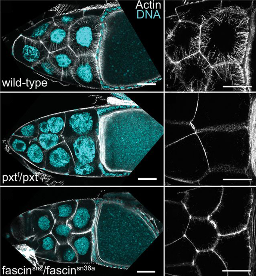
Prostaglandins (PGs) are lipid signaling molecules that regulate a wide range of processes, such as inflammation, pain, and hormone regulation. One target of PGs is the actin cytoskeleton, but how PGs affect actin filament polymerization and dynamics is not well understood. A recent paper uses the fruit fly egg chamber as a great model of how PGs regulate actin. In the fruit fly egg chamber, many nurse cells surround the oocyte (developing egg). These nurse cells are squeezed in order to dump all of their cytoplasmic contents through ring canals and into the growing egg. During this process, the nuclei of nurse cells are held in place by bundles of actin filaments to ensure that the nuclei don’t plug up the ring canals and block the transfer of the cytoplasm. PGs are an important part of this process, and they interact with an actin-bundling protein called Fascin, according to a recent paper by Groen and colleagues. Inhibition of either Fascin or Pxt, an enzyme required for PG production, results in the failure of actin bundle formation and nurse cell dumping. The loss of Fascin enhances the effects of Pxt reduction, and the overexpression of fascin suppresses the defects seen in flies with Pxt loss. Groen and colleagues show that PGs regulate Fascin, which modulates actin cytoskeleton rearrangements during nurse cell dumping. In the images above, control egg chambers (top) show parallel actin bundles (white) within nurse cells, which extend from the cell membranes to the nuclei (blue). pxt and fascin mutants (middle, bottom), however, contain little to no actin bundles in nurse cells.


ResearchBlogging.orgGroen, C., Spracklen, A., Fagan, T., & Tootle, T. (2012). Drosophila Fascin is a novel downstream target of prostaglandin signaling during actin remodeling Molecular Biology of the Cell, 23 (23), 4567-4578 DOI: 10.1091/mbc.E12-05-0417

November 27, 2012


We have a ton of neurons. And each of those neurons has many dendrites. And each dendrite has countless little dendritic spines. Thinking about how complex one single neuron is in order to receive a signal from another cell gives me an identity crisis. What if we are all just little dendritic spines on our universe’s neuron?! Was that stupid stampede for Walmart’s Black Friday sale worth it for those folks? Was my self-restraint when faced with leftover pumpkin pie worth it? Is any of it worth it?!

Dendritic spines are small actin-rich protrusions on a neuron’s dendrite, the structure that receives information from other neurons. The morphology and density of the dendritic spines can be regulated by neurotransmitters, actin dynamics, and actin-regulating proteins. The neuron-specific actin regulator cortactin-binding protein 2 (CTTNBP2) regulates the formation and maintenance of dendritic spines, and is even associated with autism spectrum disorder. A recent paper investigates the role of a CTTNBP2 homologue, CTTNBP2NL (CTTNBP2 N-terminal-like protein). Chen and colleagues found that while CTTNBP2 expression is found in the brain, CTTNBP2NL is not. In addition, CTTNBP2NL does not appear to play a role in dendritic spine formation. Although both CTTNBP2 and CTTNBP2NL associate with cortactin, a well-studied actin regulator, CTTNBP2 is associated with the cell’s cortex while CTTNBP2NL is found on actin stress fibers. In addition, Chen and colleagues found a link between CTTNBP2 and the protein phosphatase 2A (PP2A) complex, specifically with CTTNBP2 targeting the PP2A complex to dendritic spines. In the images above, cells show labels for cortactin (red) and actin fibers (blue). CTTNBP2NL (green, top) associates with stress fibers (arrows), while CTTNBP2 (green, bottom) is distributed around the cortex (arrowheads).

ResearchBlogging.orgChen, Y., Chen, C., Hu, H., & Hsueh, Y. (2012). CTTNBP2, but not CTTNBP2NL, regulates dendritic spinogenesis and synaptic distribution of the striatin-PP2A complex Molecular Biology of the Cell, 23 (22), 4383-4392 DOI: 10.1091/mbc.E12-05-0365

October 16, 2012

Molecular switches are an elegant way for a cell to regulate countless processes with a limited number of proteins. The layers of regulation that drive switch activation or inactivation allow the cell to drive one very specific process, despite the presence of all of the tools and materials to drive a thousand other processes. Today’s image is from a paper that describes one layer of regulation on a well-studied (and well-loved) set of molecular switches.

Rho GTPases are actin regulators that switch between active and inactive states. Their activation state is regulated by proteins (GEFs) that drive the GTPases into their active state, and others (GAPs) that drive them into their inactive state. RhoGDIs (Rho guanine nucleotide dissociation inhibitors) provide an additional level of control over Rho GTPase activity by sequestering Rho GTPases in inactive complexes. The selective dissociation of individual Rho GTPases from these complexes provides the cell with a context-specific response based on the actin-based structures required. A recent paper describes the function of a protein called diacylglycerol kinase ζ (DGKζ) in the release of two Rho GTPases, Rac1 and RhoA, from RhoGDI complexes. Rac1 regulates membrane ruffling and lamellipodia formation, while RhoA regulates stress fiber and focal adhesion formation. Ard and colleagues found that DGKζ-deficient cells showed signs of faulty RhoA signaling, as seen in the images above. Actin stress fibers (left column, green in merged) and focal adhesions (middle column, red in merged) appear normal in wild-type cells (top). In DGKζ-null cells (bottom row), actin stress fibers appeared condensed (arrow) and less organized, while focal adhesion distribution was impaired.

ResearchBlogging.orgArd, R., Mulatz, K., Abramovici, H., Maillet, J., Fottinger, A., Foley, T., Byham, M., Iqbal, T., Yoneda, A., Couchman, J., Parks, R., & Gee, S. (2012). Diacylglycerol kinase regulates RhoA activation via a kinase-independent scaffolding mechanism Molecular Biology of the Cell, 23 (20), 4008-4019 DOI: 10.1091/mbc.E12-01-0026