Showing posts with label cancer. Show all posts

February 12, 2015

Biologists have to wear many hats, and one under-appreciated hat is that of marketing executive. You have to properly name whatever process/protein/structure you just identified so it will be easily remembered. Whoever coined the term “invadopodia” was spot-on….the term is informative, catchy, and ignites my imagination of what it’s like inside a cell. Today’s image is from a fascinating paper on invadopodia formation.

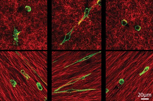
Invadopodia are dynamic protrusions of plasma membrane that locally degrade a cell’s underlying extracellular matrix (ECM). A tumor cell’s invadopodia mediate the invasion of tissue and metastasis. A recent paper describes a study of invadopodia formation within the context of a highly-concentrated collagen matrix, to better mimic the ECM of cancerous tissue. This dense collagen network, Artym and colleagues found, triggers robust invadopodia formation and ECM degradation, in both cancerous and non-cancerous cell lines. This invadopodia formation did not require altered gene or protein expression, but did require phosphorylation of kindlin2, part of a complex integrin regulatory network. As seen in the images, the high-density fibrillary collagen (HDFC, top) network triggered the induction of many more invadopodia (yellow dots) than a gelatin-based matrix (bottom).  

Artym, V., Swatkoski, S., Matsumoto, K., Campbell, C., Petrie, R., Dimitriadis, E., Li, X., Mueller, S., Bugge, T., Gucek, M., & Yamada, K. (2015). Dense fibrillar collagen is a potent inducer of invadopodia via a specific signaling network originally published in the Journal of Cell Biology, 208 (3), 331-350 DOI: 10.1083/jcb.201405099

November 19, 2014

You might not be able to get rid of the bad guys, but you can still win the battle if you cripple their mobility. Today’s image is from a paper describing how a tumor’s microenvironment can predict the motility of individual tumor cells.

Metastasis is the spread of cancer cells throughout the body. The motility of tumor cells depends on the microenvironment around them, and a recent paper systematically looks at how that microenvironment can predict or alter the behavior of tumor cells. Gligorijevic and colleagues tracked the motility of individual mouse mammary carcinoma cells in vivo using high-resolution multi-photon microscopy, and found that tumor cells exhibited either fast or slow locomotion. Those tumor cells with slow locomotion also exhibited invadopodia, protrusions that Gligorijevic and colleagues directly link to degradation of the underlying extracellular matrix and metastasis. While no single parameter of the tumor’s microenvironment could predict the locomotion of tumor cells, a support vector machine algorithm indicated how combinations of many parameters could predict tumor cell phenotype and behavior. By characterizing the heterogeneous microenvironment of a tumor and predicting the location and behavior of metastatic tumor cells, researchers can better understand treatment of tumors and the varying responses. Images above show protrusions (arrowheads, over 30 minutes) on two different tumor cells with slow locomotion, with protrusions facing collagen fibers (purple).

BONUS!! Check out a movie of these protrusions below. Note that some protrusion face collagen fibers (purple, panels a and b), and some protrude into blood vessels (red, panels c and d).

BONUS!! Check out Bojana Gligorijevic ‘s interview with SciArt about her images, research, and art here.

Gligorijevic, B., Bergman, A., & Condeelis, J. (2014). Multiparametric Classification Links Tumor Microenvironments with Tumor Cell Phenotype PLoS Biology, 12 (11) DOI: 10.1371/journal.pbio.1001995

September 25, 2014

While taking an awesome cell biology course in college, I was coming to terms with my mother’s recent ovarian cancer diagnosis. The scientist in my head couldn’t shake the curiosity about how my mother’s cells could have betrayed her so royally. This intersection of basic cell biology and cancer kick-started my interest in cell biology research. Today’s image is from a paper showing a role for the ARF tumor suppressor in maintaining chromosomal stability. THIS paper is one of the million billion reasons why basic research is necessary and important. 

The ARF tumor suppressor is mutated or absent in many cancers, and is known to stabilize p53 in response to cellular stress. Other, p53-independent roles for ARF contribute to its role as a tumor suppressor, but are not well understood. A recent paper describes ARF’s function in chromosome segregation during mitosis, via Aurora B regulation. Britigan and colleagues show that loss of ARF results in aneuploidy, or an incorrect number of chromosomes, stemming from chromosome segregation and spindle organization defects. These defects can be rescued through overexpression of the Aurora B kinase, which helps ensure proper kinetochore-spindle interactions and is overexpressed in some cancers. In the images above, ARF-/- cells (right column) show defects throughout mitosis, when compared to normal cells (left). Defects include misaligned chromosomes during metaphase (top, middle rows), and lagging chromosomes during anaphase (bottom).

Britigan, E., Wan, J., Zasadil, L., Ryan, S., & Weaver, B. (2014). The ARF tumor suppressor prevents chromosomal instability and ensures mitotic checkpoint fidelity through regulation of Aurora B Molecular Biology of the Cell, 25 (18), 2761-2773 DOI: 10.1091/mbc.E14-05-0966

January 22, 2014

The C-word is a dirty, dirty word that every single person dreads hearing. Cancer touches every family at some point (or so it seems), so the effort put forth to finding a “cure” for cancer is huge. Today’s image is from a very exciting paper that shows how specific cells in a breast tumor lead the charge towards invasion. 

The spread of cancer, or metastasis, can occur either through the invasion of single tumor cells into nearby tissue or by collective invasion of several cells as a cohesive unit. A recent paper from Andy Ewald’s lab at Johns Hopkins describes the identification of cells involved in collective invasion, using a 3D assay of primary breast tumors invading other tissue. Kevin Cheung and colleagues found that in mouse breast cancer models and diverse human breast tumors, the cells leading the invasion charge are distinct from the bulk tumor cells, and express the basal epithelial genes cytokeratin-14 (K14) and p63. Additionally, knockdown of either K14 or p63 could block collective invasion in advanced carcinomas. In the images above, leading invasive cells express K14 (middle image, green) and are distinct from the bulk of the mouse mammary tumor.

BONUS!! For a great “Out of the Box” description of these very cool results, click here.

Kevin J. Cheung, Edward Gabrielson, Zena Werb, Andrew J. Ewald (2013).  Collective Invasion in Breast Cancer Requires a Conserved Basal Epithelial Program.  Cell, 15 (7), 1639–1651. http://dx.doi.org/10.1016/j.cell.2013.11.029  Copyright ©2013 Elsevier Ltd. All rights reserved.

March 19, 2013

After a lovely Spring Break with my family, HighMag is back in action.  As an oldie, Spring Break takes on a whole new meaning than it did years ago…this pregnant lady didn’t have one single beer!

 Cancer is a series of cellular mistakes, mistakes that are too far gone to fix without the help of the medical field.  Understanding the mistakes at the most basic cellular level is key to fighting the war on cancer, and a recent paper is a fine example of this.

A great model for understanding cell polarity is the intestinal epithelial sheet of cells that provides a barrier between the inside of the intestine and the body.  The different polarized domains – apical and basal – each have a discrete set of adhesion and membrane proteins trafficked to them.  One membrane trafficking protein, Rab25, is a tumor suppressor for colon cancer in both humans and mice.  A recent paper investigates the link between Rab25 and the polarized intestinal cells involved in colon cancer.  Krishnan and colleagues found that a reduction of Rab25 levels in cultured endothelial cells resulted in increased cell invasion and a loss of certain integrins, adhesion proteins, at the plasma membrane.  Rab25 loss also affected the transcription of several genes including the transcription factor ETV4, suggesting that Rab25’s effect on cell polarity is through gene regulation.  The scanning electron images above show brush border microvilli, the fingerlike-projections seen in intestinal epithelial sheets that serve to increase absorption of nutrients from the intestine.  In Rab25-reduced cells (middle row), the brush border is sparsely-packed and disorganized when compared to control cells (top row).  Bottom row shows Rab25-reduced cells in which rabbit Rab25 was reintroduced, and the rescued brush border is more organized.

ResearchBlogging.orgKrishnan M, Lapierre LA, Knowles BC, & Goldenring JR (2013). Rab25 regulates integrin expression in polarized colonic epithelial cells. Molecular biology of the cell, 24 (6), 818-31 PMID: 23345591

March 4, 2013

To me, science is a world of optimism.  Researchers are constantly looking for cures for countless diseases, and with intelligence, a bit of luck, and grant funding their hard work rolls into results that may lead to new therapies.  I always look forward to seeing how some of the most basic cell biology experiments can lead to something big, and I again fall in love with the whole process.

Medulloblastoma is the most common pediatric brain tumor, yet the treatments are highly toxic and are associated with high morbidity.  A recent paper found that PIGF (placental growth factor) is expressed in most medulloblastomas, regardless of their subtype, and that high expression of the PIGF receptor Nrp1 is associated with poor survival of patients.  Snuderl and colleagues then showed that growth and spread of medulloblastomas are dependent on PIGF and Nrp1, suggesting that PIGF and Nrp1 could serve as targets for future therapies for medulloblastoma.  In the two-photon microscopy images above, the vasculature (green) of medulloblastomas (blue) was less dense in tumors treated with a PIGF-blocking antibody (bottom), compared with control (top) over time, during development.  In the antibody-treated tissue, there was even some vessel regression (white arrows, bottom).

ResearchBlogging.orgSnuderl, M., Batista, A., Kirkpatrick, N., Ruiz de Almodovar, C., Riedemann, L., Walsh, E., Anolik, R., Huang, Y., Martin, J., Kamoun, W., Knevels, E., Schmidt, T., Farrar, C., Vakoc, B., Mohan, N., Chung, E., Roberge, S., Peterson, T., Bais, C., Zhelyazkova, B., Yip, S., Hasselblatt, M., Rossig, C., Niemeyer, E., Ferrara, N., Klagsbrun, M., Duda, D., Fukumura, D., Xu, L., Carmeliet, P., & Jain, R. (2013). Targeting Placental Growth Factor/Neuropilin 1 Pathway Inhibits Growth and Spread of Medulloblastoma Cell, 152 (5), 1065-1076 DOI: 10.1016/j.cell.2013.01.036
Copyright ©2013 Elsevier Ltd. All rights reserved.

October 23, 2012

Understanding how a cell works normally is hard enough for biologists. Understanding how a cancerous cell works is exponentially harder—there are different stages of tumorigenesis and countless different types of cancer and countless different environments within the body. Today’s image is from a study that takes a systematic approach to understanding the interactions between cancerous cells and their environment.

The progression of tumor cells to metastatic cancer cells correlates with poor prognoses for cancer patients. The steps that drive cancer cells to spread (metastasis) are not well understood, but may be effective targets for chemotherapies. For example, tumor cells lose adhesion to their underlying extracellular matrix (ECM) prior to spreading to other regions of the body. Understanding this loss of cell-ECM adhesion may guide the development of new therapies. A recent paper describes the systematic analysis of cell-ECM adhesion in tumor cells by using robotically spotted arrays of 768 paired combinations of ECM molecules. Reticker-Flynn and colleagues monitored the adhesion profiles of lung cancer cell lines at different stages of cancer progression on these arrays of ECM molecules, and found ECM-cell interactions that may be successful therapeutic targets. The images above show an array of spotted ECM protein combinations (visible through immuno- and fluorescent-labeling, top), and examples of cells adhered to the ECM spots (bottom images).

ResearchBlogging.orgReticker-Flynn, N., Malta, D., Winslow, M., Lamar, J., Xu, M., Underhill, G., Hynes, R., Jacks, T., & Bhatia, S. (2012). A combinatorial extracellular matrix platform identifies cell-extracellular matrix interactions that correlate with metastasis Nature Communications, 3 DOI: 10.1038/ncomms2128
 Adapted by permission from Macmillan Publishers Ltd, copyright ©2012

September 13, 2012


I’m sure you've been here before….you’re at a family gathering, and some distant relative or in-law hears that your research involves worms, flies, or yeast. You are snidely asked what use it is to do research on that, and then asked what kind of job you could actually get with that kind of background. This happened to me (at a funeral), but I didn’t have either the speed or the smugness to rattle off the list of diseases understood or medications developed thanks to these kinds of organisms. Next time, I’ll just pass out a copy of the paper that today’s image comes from—booyah!

Throughout evolution, many genes are coopted for use in diverse organisms. Recently, the gene network that maintains the cell wall in yeast, a fungus, was discovered to also play a role in vertebrate angiogenesis. Angiogenesis is the growth of blood vessels from pre-existing vessels, and is an important step in transforming a tumor into a spreading, malignant cancer. The same research group that realized this yeast-angiogenesis link suggested that drugs affecting the yeast cell wall may also function as angiogenesis inhibitors for chemotherapy. Cha and colleagues found that an inexpensive antifungal drug called thiabendazole could block angiogenesis in animal models and human cells. Specifically, the drug disassembles newly-sprouted blood vessels. When Cha and colleagues grafted human tumors into mice, they found that thiabendazole treatment slowed tumor growth and limited growth of the vascular network. In the images above, the network of blood vessels in a Xenopus frog embryo is disrupted after thiabendazole treatment (bottom), compared to a wild type embryo (top).

ResearchBlogging.orgCha HJ, Byrom M, Mead PE, Ellington AD, Wallingford JB, & Marcotte EM (2012). Evolutionarily repurposed networks reveal the well-known antifungal drug thiabendazole to be a novel vascular disrupting agent. PLoS biology, 10 (8) PMID: 22927795

March 5, 2012

Scenario: You’re at my house for a dinner and I make you a mind-blowing chocolate tart. You ask why it is so durn good, and I pass along the fact that Nutella is the ass-kicking ingredient. Next thing you know, you’re trying to add Nutella to everything at home….and by golly, it makes (many) things taste better. Today’s image is from a paper characterizing the relationship between the many cellular changes after treatment by antimitotic drugs. Knowing how these drugs work (by analogy, finding the ass-kicking “ingredient”) can help folks develop improved anti-cancer drugs.

Many anticancer drugs are antimitotic drugs, meaning they function by blocking the progress of cell division during mitosis. In addition to arresting mitosis, these drugs cause apoptosis, DNA damage, and induction of p53 (a tumor suppressor gene), and a recent paper investigates the relationship between all of these events. After treating cells with powerful antimitotic drugs, Orth and colleagues found that the resulting prolonged mitotic arrest (or slippage from that arrest) causes DNA damage, which in turn causes an induction of p53. The DNA damage was inhibited when these treated cells were prevented from launching the pathway for apoptosis, which is programmed cell death. So, Orth and colleagues concluded that the prolonged mitotic arrest caused by antimitotic drugs results in a partial activation of apoptosis. Understanding this partial apoptotic response in the context of treating tumors should help guide development of improved cancer therapies. The images above show increasing DNA damage (red spots) after prolonged treatment with an antimitotic drug, compared with an untreated cell (top left). By 16 and 48 hours of drug treatment (bottom), cells had very high levels of DNA damage. Arrows point to mitotic cells.

ResearchBlogging.orgOrth, J., Loewer, A., Lahav, G., & Mitchison, T. (2011). Prolonged mitotic arrest triggers partial activation of apoptosis, resulting in DNA damage and p53 induction Molecular Biology of the Cell, 23 (4), 567-576 DOI: 10.1091/mbc.E11-09-0781

March 1, 2012

Tumors begin when a cell goes rogue. These rogue cells turn against us in a most unforgiving way, even when their environment and neighboring cells try to put the kibosh on such behavior. Today’s image is from a fascinating paper describing how a single mutant cell in a highly organized environment can move out of the tissue, similar to what is seen in some tumors.

The development of a tumor begins with sporadic mutations of oncogenes, but these mutant cells are frequently in a highly organized tissue that limits its growth and movement, two key elements to the spread of cancer. A recent paper describes how single mutant cells are able to overcome their suppressive environment and move out of the tissue. To study this, Leung and Brugge grew mammalian cells in 3D cultures and followed clusters of cells with hollow lumens, called acini, after overexpressing different oncogenes. The overexpression of ERBB2, an oncogene overexpressed in 30% of breast tumors, caused outgrowth of the mutated cells from the cluster’s epithelial layer and into the lumen, a feature commonly seen in carcinoma in situ (non-invasive) breast tumors. In addition, Leung and Brugge found that this is a highly regulated process, as the mutation also causes changes in adhesion between the cell and its underlying matrix. These changes also support the survival and growth of the mutant cells. As seen in the images above, control cells (green, top) remained within the highly organized epithelial layer of the cluster over a course of 56 hours, but a single ERBB2-overexpressing cell (green, bottom) dissociated from the epithelial layer and moved into the hollow lumen.

ResearchBlogging.orgLeung, C., & Brugge, J. (2012). Outgrowth of single oncogene-expressing cells from suppressive epithelial environments Nature, 482 (7385), 410-413 DOI: 10.1038/nature10826
Adapted by permission from Macmillan Publishers Ltd, copyright ©2012

August 15, 2011

You may live in many places throughout your life, but you have only one hometown. No matter what, your core is deeply affected by where you grew up (for me, that core is made of a fondness for fries covered with gravy, impassioned shouts of Bruuuuuce, and traumatic experiences with a teasing comb and hairspray…yes, that’s New Jersey). Just like us, a tumor is affected by its origin…its growth, malignancy, and responsiveness to treatment are all dependent on where the cancer cells came from. A recent paper tracks tumor growth to determine the origin of a certain type of tumor.

The countless types of cancer each come from different cell types. A tumor’s potential for growth, spreading, and treatment are heavily dependent on the tumor’s cell of origin. Malignant glioma is a deadly type of brain tumor, and a recent paper has determined the cell of origin for this cancer. Lui and colleagues used a technique called MADM (mosaic analysis with double markers) that mimics the genetic mutations seen in gliomas. This technique labels mutant cells green and normal cells red, enabling them to track how and when the mutant cells develop into tumors. These biologists started out with neural stem cells, which were suspected as the cells of origin for gliomas, yet found that cells called oligodendrocyte precursor cells (OPCs) are the cells of origin. The images above show mutant MADM neurons. Left and right images show MADM labeling of mutant (green) and normal (red) cells, while middle image shows the nuclei of cells (blue in merged).

ResearchBlogging.orgLiu, C., Sage, J., Miller, M., Verhaak, R., Hippenmeyer, S., Vogel, H., Foreman, O., Bronson, R., Nishiyama, A., Luo, L., & Zong, H. (2011). Mosaic Analysis with Double Markers Reveals Tumor Cell of Origin in Glioma Cell, 146 (2), 209-221 DOI: 10.1016/j.cell.2011.06.014
Copyright ©2011 Elsevier Ltd. All rights reserved.

July 25, 2011


They invade...they proliferate...they destroy. It sounds like the tagline for a terrible summer blockbuster starring Samuel L. Jackson and an animated sidekick voiced by one of the Kardashians, but it’s the tagline of something far more sinister and real. I’m talking about tumors. Today’s image is from a paper showing how a membrane protein called caveolin-1 can support tumor invasion.

Caveolin-1 is a membrane protein and a major component of caveolae, which are small membrane invaginations that participate in endocytosis. A recent paper finds that caveolin-1 also functions in cell elongation, migration, and invasion by remodeling a cell’s microenvironment (aka “stroma”). Specifically, Goetz and colleagues found that caveolin-1 affects stromal architecture by regulating the activity of Rho GTPase, a signaling protein frequently involved in actin dynamics. This caveolin-1-inspired remodeling of the stroma is significant for tumor biology, too—the stiffness, contractility, and general architecture of a tumor’s stroma can affect its growth, invasion, and metastasis. In the images above, tumor cells (green) were cultured in a 3D-gels with fibroblast cells (red) that expressed caveolin-1 (top row) or did not express caveolin-1(bottom row). When tumor cells were surrounded by caveolin-1-expressing cells, they were able to invade further into the gel.

ResearchBlogging.orgGoetz, J., Minguet, S., Navarro-Lérida, I., Lazcano, J., Samaniego, R., Calvo, E., Tello, M., Osteso-Ibáñez, T., Pellinen, T., Echarri, A., Cerezo, A., Klein-Szanto, A., Garcia, R., Keely, P., Sánchez-Mateos, P., Cukierman, E., & Del Pozo, M. (2011). Biomechanical Remodeling of the Microenvironment by Stromal Caveolin-1 Favors Tumor Invasion and Metastasis Cell, 146 (1), 148-163 DOI: 10.1016/j.cell.2011.05.040
Copyright ©2011 Elsevier Ltd. All rights reserved.

June 30, 2011

Between the high number of breast cancer patients and the pink ribbons seen all over, breast health is and will always be a hot topic. The breast is a fascinating system of different cell and tissue types, and today’s image is from a paper looking at a population of epithelial cells in the breast.

Breast tissue contains two layers of epithelial sheets—an outer layer of myoepithelial cells (MECS) and an inner layer of luminal epithelial cells (LECs). The LECs greatly expand during certain events, such as pregnancy and tumorigenesis, which results in either thinner or discontinuous coverage by the outer layer of MECs. Because of this, the MEC layer serves as an epithelial “gatekeeper,” by generating boundaries that help organize breast tissue. A recent paper looks at this gatekeeper function of MECs and finds that two proteins, SLIT and ROBO2, are important in regulating the proliferation of MECs. And, in turn, the growth of the MEC layer regulates the branching of mammary tissue. Image above shows mammary tissue in normal (left) or Robo mutant (right) mice. The loss of Robo leads to excessive branching (close-up views of boxed regions on bottom).

ResearchBlogging.orgMacias, H., Moran, A., Samara, Y., Moreno, M., Compton, J., Harburg, G., Strickland, P., & Hinck, L. (2011). SLIT/ROBO1 Signaling Suppresses Mammary Branching Morphogenesis by Limiting Basal Cell Number Developmental Cell, 20 (6), 827-840 DOI: 10.1016/j.devcel.2011.05.012
Copyright ©2011 Elsevier Ltd. All rights reserved.

March 24, 2011

Cancer cells have taught biologists about a lot of wacky things that can happen when things don’t go merrily along for a cell. Entosis is a process in which a living cell is internalized into a neighboring cell, and has been found to occur in some tumors. A recent paper describes exactly what can go wrong here.

Aneuploidy refers to a cell having an incorrect number of chromosomes, and is a feature of many cancers. Typically, aneuploidy occurs from a failure in cytokinesis, the physical division of a cell after mitosis, due to misregulation or mutation of genes involved in cell division. Sometimes, however, aneuploidy can occur from a non-genetic failure of cytokinesis, according to a recent paper. In this paper, Krajcovic and colleagues look at cytokinesis failures due to entosis, a process in which living cells are internalized by their neighboring cells. These cell-in-cell structures are found in some tumors, and the outer “host” cell is frequently aneuploid. This aneuploidy occurs when the internalized cell physically disrupts the constriction required to cleave two cells during cytokinesis, as seen in the images above. Cytokinesis of the cell-in-cell structure (left) is not going well compared with a normal cell (right). Red labels (and in black and white insets) mark active constriction during cytokinesis, and should be symmetric around the cells. The mitotic spindle is labeled in green, and chromosomes in blue.


BONUS!! Movie of above cell, attempting cytokinesis, can be found here. More cool movies from this paper can be found here.

ResearchBlogging.orgKrajcovic, M., Johnson, N., Sun, Q., Normand, G., Hoover, N., Yao, E., Richardson, A., King, R., Cibas, E., Schnitt, S., Brugge, J., & Overholtzer, M. (2011). A non-genetic route to aneuploidy in human cancers Nature Cell Biology, 13 (3), 324-330 DOI: 10.1038/ncb2174
Adapted by permission from Macmillan Publishers Ltd, copyright 2011

February 14, 2011

Cancer cells are smart little guys, unfortunately. Many cancer therapies target a specific type of cancer cell migration, but many cancer cells are able to switch to a different mode of migration and evade the attack. Today’s image is from a paper looking at these two modes of migration by using 3D cultures of malignant breast cancer cells.

Malignant cancer cells are able to spread beyond the initial tumor, and their migration can occur from using either of two types of cancer cell migration—mesenchymal and amoeboid. Mesenchymal migration is characterized by elongated cancer cells that can undergo proteolysis of and adhesion to the extracellular matrix on which it is migrating. Cancer cells undergoing amoeboid migration are rounded and squeeze through extracellular spaces without proteolysis of or adhesion to the matrix. A recent paper looks at the distinct roles of two different adhesion adaptor proteins – paxillin and Hic-5 – in these two modes of migration. Images above are of breast cancer cells (green) grown in two different types of 3D cultures (top and bottom). Compared with the mixed morphologies of control breast cancer cells (left images), cells without paxillin (middle images) had elongated, mesenchymal morphologies. Breast cancer cells without Hic-5 (right images) had more rounded, amoeboid morphologies.

ResearchBlogging.orgDeakin, N., & Turner, C. (2010). Distinct roles for paxillin and Hic-5 in regulating breast cancer cell morphology, invasion, and metastasis Molecular Biology of the Cell, 22 (3), 327-341 DOI: 10.1091/mbc.E10-09-0790

February 7, 2011

Cancer is not a disease…it is many many diseases. Some cancers come about gradually, while others can occur from a single catastrophic cellular event, according to a recent paper.

Most cancers are believed to progress through a series of genetic changes that gradually allows cells to become cancerous and spread. These genetic changes are random, but can be pushed along by carcinogens or DNA repair problems. Sometimes, though, there can be big pulses of genetic change that encourage cancer progression, and a recent paper describes how single catastrophic cellular events can cause major genomic rearrangements that quickly lead to cancer. In this paper, Stephens and colleagues found multiple cancer samples with tens to hundreds of genomic rearrangements that were caused by a single catastrophic event, and termed this phenomenon “chromothripsis.” Evidence of chromothripsis can be found in at least 2-3% of all cancers, including many subtypes, and in about 25% of bone cancers. Images above show chromosomes from a renal cancer cell line, with chromosome 5 marked with different fluorescent probes that find specific genomic regions. Compared to the normal chromosome 5, the derivative chromosome 5 has gross rearrangements suggesting chromothripsis, as seen by the close juxtaposition of all of the fluorescent probes.

ResearchBlogging.orgStephens, P., Greenman, C., Fu, B., Yang, F., Bignell, G., Mudie, L., Pleasance, E., Lau, K., Beare, D., & Stebbings, L. (2011). Massive Genomic Rearrangement Acquired in a Single Catastrophic Event during Cancer Development Cell, 144 (1), 27-40 DOI: 10.1016/j.cell.2010.11.055
©2011 Elsevier Ltd. All rights reserved.

January 10, 2011

My last blog post was about how the immune system gets rid of invaders, but this post is about a paper showing some fascinating results about immune cells actually helping an unwanted resident, a transformed cell. This paper provides a very cool addition to the idea that some cancers look like non-healing wounds.

Zebrafish are a very powerful organism to use in the lab for many reasons, one of which is their transparency during development. Feng and colleagues recently took advantage of this in order to find and image the interactions between immune cells and oncogene-transformed cells as they initiate cancerous growth. These transformed cells recruit leukocytes, which are white blood cells, using H2O2 in a similar process that wounds use to recruit immune cells as part of the inflammatory response. When H2O2 synthesis was blocked, leukocytes were not recruited to the transformed cells and the number of transformed cells was reduced. The authors’ results suggest that the interactions with leukocytes serve to support proliferation of the transformed cells. Image above shows leukocytes (red) and transformed cells (green) interacting by forming tethers between the two cells.

BONUS!! Very cool video of a tether forming between the cell types can be seen here. And, more cool videos from this paper can be found here.

ResearchBlogging.orgFeng, Y., Santoriello, C., Mione, M., Hurlstone, A., & Martin, P. (2010). Live Imaging of Innate Immune Cell Sensing of Transformed Cells in Zebrafish Larvae: Parallels between Tumor Initiation and Wound Inflammation PLoS Biology, 8 (12) DOI: 10.1371/journal.pbio.1000562

January 3, 2011

Cancer is a loaded word for many biologists—it is made up of thousands of different diseases when you realize how many different paths can be taken in order for cells to lead to cancer. There are so many biologists investigating cancer, and sometimes there are discoveries that shake up our understanding. These shake-ups are key to making the big steps towards a cure that patients, survivors, and victims all hope for.

Cancer progression involves growth of a tumor as well as metastatic spread of cancerous cells to other tissues. Tumor growth requires the development of a blood supply for the tumor, and it was previously known that outside blood vessels get induced to sprout new vessels at the site of the tumor. A recent set of papers in Nature describe the ability of tumor stem cells in glioblastoma cancer to induce production of endothelial cells used in vessel formation. Images above show endothelial glioblastoma cells forming tubular vascular networks, a key step towards vessel formation, in a three-dimensional culture. DAPI shows nuclei of cells (blue), CD105 indicates dividing angiogenic endothelial cells (green), and DiI-AcLDL labels vascular endothelial cells (red). Image on right shows phase contrast of the tubular network.


ResearchBlogging.orgAdapted by permission from Macmillan Publishers Ltd, copyright 2010.
Wang, R., Chadalavada, K., Wilshire, J., Kowalik, U., Hovinga, K., Geber, A., Fligelman, B., Leversha, M., Brennan, C., & Tabar, V. (2010). Glioblastoma stem-like cells give rise to tumour endothelium Nature, 468 (7325), 829-833 DOI: 10.1038/nature09624


Accompanying paper:
Ricci-Vitiani, L., Pallini, R., Biffoni, M., Todaro, M., Invernici, G., Cenci, T., Maira, G., Parati, E., Stassi, G., Larocca, L., & De Maria, R. (2010). Tumour vascularization via endothelial differentiation of glioblastoma stem-like cells Nature, 468 (7325), 824-828 DOI: 10.1038/nature09557

Fantastic News and Views paper on these results:
Bautch, V. (2010). Cancer: Tumour stem cells switch sides Nature, 468 (7325), 770-771 DOI: 10.1038/468770a