Showing posts with label myosin. Show all posts

December 18, 2014


You might not want the dreaded tube socks in your Christmas stocking this year, but you do appreciate the actual tubes that your body depends on in just about every organ system. A recent paper in PLOS Biology describes tube formation in the fly renal system and the signals that regulate it.

Tubes generally start as buds that dramatically elongate during development, but the cell rearrangements that occur during tubulogenesis are not completely understood. Saxena and colleagues recently used the developing fly renal system to track cell movements during tube formation. Tubule elongation primarily occurs through convergent extension, during which cells intercalate along the length of the tube. During these rearrangements, the number of cells around the circumference of the tube drops as the number of cells along the tube increases. Saxena and colleagues show that epidermal growth factor localized at the tip cells of the distal end of the tube guides the polarity of cell rearrangements, via polarization of Myosin II within individual cells. Finally, without proper tube elongation, animals have abnormal excretory function and osmoregulation, leading to lethality. In the images above, the top row shows failure of tube elongation after laser ablation of the distal tip cells (arrowheads). Bottom row shows normal tube elongation without laser ablation of tip cells (arrowheads).

Saxena, A., Denholm, B., Bunt, S., Bischoff, M., VijayRaghavan, K., & Skaer, H. (2014). Epidermal Growth Factor Signalling Controls Myosin II Planar Polarity to Orchestrate Convergent Extension Movements during Drosophila Tubulogenesis PLoS Biology, 12 (12) DOI: 10.1371/journal.pbio.1002013

October 28, 2014

If you’ve ever tried to get your kids to share a donut, you understand the importance to dividing things equally (and learning crucial lessons…just buy more donuts next time...I mean, seriously!). Cell division is no different—chromosomes and organelles must all get divided equally. Today’s images are from a paper showing how mitochondria are positioned during cell division in order to allow equal segregation.

Many years of research have focused on the equal segregation of chromosomes during cell division. Organelles such as mitochondria must also be segregated equally in a dividing cell, and errors in this process can lead to disease. A recent paper identifies the actin motor Myosin-XIX (Myo19) as a key player in mitochondrial partitioning during cell division. Myo19 is localized to mitochondria, and cells depleted of Myo19 have an abnormal distribution of mitochondria. Rohn and colleagues found that cells lacking Myo19 experience stochastic division failure, suggesting that mitochondria are physically preventing successful cell division. The images above show dividing cells labeled to visualize mitochondria (green) and the mitotic spindle (red) in control cells (top two rows) and cells depleted of Myo19 (bottom two rows). Without Myo19, mitochondria moved towards spindle poles at the onset of anaphase, causing an asymmetric distribution at division when compared with control cells.

BONUS!! Here is a rotating 3D reconstruction of an A549 stained to visualize microtubules (green), mitochondria (red), and DNA (blue). Omar Quintero, HighMag friend and a co-author from today’s paper, loves this image: “I like it because it reminds me of the scenes in StarWars where the Rebels are planning their attack on the Death Star.”

Rohn, J., Patel, J., Neumann, B., Bulkescher, J., Mchedlishvili, N., McMullan, R., Quintero, O., Ellenberg, J., & Baum, B. (2014). Myo19 Ensures Symmetric Partitioning of Mitochondria and Coupling of Mitochondrial Segregation to Cell Division Current Biology DOI: 10.1016/j.cub.2014.09.045

Copyright ©2014 Elsevier Ltd. All rights reserved.

April 17, 2014

The endoplasmic reticulum and humans have quite a bit in common. Both are dynamic and constantly changing, but both also need something to ground and stabilize them. Maybe I’m reading too much into the beauty of the ER, but the image today is from a paper that only fuels my fascination.

The endoplasmic reticulum (ER) is a large, complex membrane-bound organelle that spreads throughout the cell and hosts the synthesis, folding, and sorting of membrane and secretory proteins. This network is dynamic and constantly rearranging, with diverse structural and functional domains. A recent paper describes the investigation into the role of the actin cytoskeleton in regulating ER sheet persistence and maintenance, which is important as the stationary domain of the ER. Joensuu and colleagues identified a subset of actin filaments associated with the ER, specifically to polygons defined by ER tubules and sheets. Actin depolymerization caused ER sheet fluctuation and resulted in a defective ER network. The actin motor myosin 1c also localizes to these actin filament arrays. In the images above, actin filament arrays (magenta, arrows) localize to the polygons (asterisks) associated with the ER (green).

Joensuu, M., Belevich, I., Ramo, O., Nevzorov, I., Vihinen, H., Puhka, M., Witkos, T., Lowe, M., Vartiainen, M., & Jokitalo, E. (2014). ER sheet persistence is coupled to myosin 1c-regulated dynamic actin filament arrays Molecular Biology of the Cell, 25 (7), 1111-1126 DOI: 10.1091/mbc.E13-12-0712

March 5, 2014

Mitochondria are the cellular power plants, but bigger power plants are not always a good thing. Defects in the regulation of mitochondrial size and dynamics can cause neurodegenerative diseases such as Alzheimer’s disease. Today’s image is from a paper describing an important player in mitochondrial division, or fission.

Mitochondria serve as the cellular power plants due to their production of ATP, the cell’s energy source, and are quite dynamic, with fusion and fission events occurring regularly. Mitochondrial fission is how mitochondria divide, but fission also plays an important role in apoptosis and ridding the cell of damaged mitochondrial components. In current models of fission, the GTPase dynamin (Drp1) forms a ring around and constricts the mitochondrial membranes. A recent paper describes the importance of the myosin II, an actin motor, in Drp1-mediated fission. Korobova and colleagues found that inhibition of myosin II resulted in abnormally long mitochondria. This inhibition of myosin II also affected the presence of Drp1 at mitochondria. In the images above, the use of blebbistatin, a myosin II chemical inhibitor, resulted in long mitochondria (right), compared to control mitochondria (left).

Korobova, F., Gauvin, T., & Higgs, H. (2014). A Role for Myosin II in Mammalian Mitochondrial Fission Current Biology, 24 (4), 409-414 DOI: 10.1016/j.cub.2013.12.032
Copyright ©2014 Elsevier Ltd. All rights reserved.

June 11, 2013

 I’m willing to bet that most scientists were pretty destructive as kids.  Not intentionally destructive, though…I bet we all liked taking things apart to see what each part of a toy did.  Maybe your Teddy Ruxpin eventually sounded like a demonic doll after your experiments, or you finally pulled apart your Etch-A-Sketch to uncover the magic.  Either way, it was early training for what scientists do every day to understand cells better.  Today’s image is from a paper that takes this route to understand cell adhesion better.

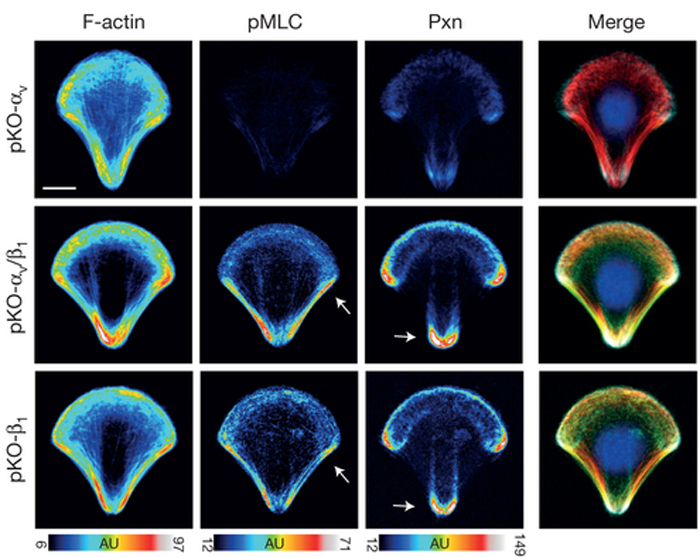
Integrins are transmembrane proteins that attach a cell to another cell or its surrounding extracellular matrix, so their role in regulating cell adhesion in normal cell function, development, and disease is of serious interest to biologists.  There are several types of integrins, frequently expressed together at the same time.  A recent paper describes a system to investigate how specific integrins function in cell adhesion, and found notable differences.  Schiller and colleagues expressed either β1- or αv-class integrins in fibroblasts lacking all other integrins, and found that β1-class integrins are important in small peripheral adhesions, while αv-class integrins are important for large focal adhesions.  In the images above, activity of the actin-based motor myosin and presence of the focal adhesion protein paxillin are shown in cells plated on micropatterned shapes.  Cells with both β1- and αv-class integrins (middle row) have the highest myosin activity (pMLC) and paxillin signal, while cells with either β1- or αv-integrin alone have lower myosin activity and paxillin signal.  

ResearchBlogging.orgSchiller, H., Hermann, M., Polleux, J., Vignaud, T., Zanivan, S., Friedel, C., Sun, Z., Raducanu, A., Gottschalk, K., Théry, M., Mann, M., & Fässler, R. (2013). β1- and αv-class integrins cooperate to regulate myosin II during rigidity sensing of fibronectin-based microenvironments Nature Cell Biology, 15 (6), 625-636 DOI: 10.1038/ncb2747
Adapted by permission from Macmillan Publishers Ltd, copyright ©2013 

May 13, 2013

Molecular motors are some of the raddest things in a cell.  They can walk along cytoskeletal elements such as microtubules and actin filaments, and the list of cellular events that they participate in is a long, long list.  Today’s image is from a paper showing a beautiful pattern of nonmuscle myosin II in epithelial cells.

Epithelial cells assemble junctions to adhere to one another, and the actin motor nonmuscle myosin II (NMII) is a major component of these epithelial apical junctions.  NMII helps the epithelial sheet respond to morphogenesis and changes in tissue homeostasis, and a recent paper describes how the network of NMII motors does so.  Ebrahim and colleagues have found that NMII in the apical junctional complex of epithelial cells assembles into precise muscle-like sarcomere units that form a belt around each cell.  The sarcomeres of neighboring cells are aligned, in turn assembling into a contractile network that can result in changes in cell shape.  In the images above, NMII (green) is seen in repeated sarcomere units around each cell (actin is in red).  NMII puncta are paired together in neighboring cells.  Arrows (middle) point to the junctions between three cells, seen at higher magnification on the right.

ResearchBlogging.orgEbrahim, S., Fujita, T., Millis, B., Kozin, E., Ma, X., Kawamoto, S., Baird, M., Davidson, M., Yonemura, S., Hisa, Y., Conti, M., Adelstein, R., Sakaguchi, H., & Kachar, B. (2013). NMII Forms a Contractile Transcellular Sarcomeric Network to Regulate Apical Cell Junctions and Tissue Geometry Current Biology, 23 (8), 731-736 DOI: 10.1016/j.cub.2013.03.039 
Copyright ©2013 Elsevier Ltd. All rights reserved.

October 1, 2012

There are a few things in our world that only turn to the right—Derek Zoolander and UPS trucks, for example. Maybe they could be inspired by the images above of myosin IC, which turns in left-hand curves.

Myosins are actin-based motors that function in countless cell processes. Class I myosins link cell membranes with the actin cytoskeleton and bind directly to lipid membranes. Within this class of myosins, myo1c is important in hearing, endocytosis, and membrane trafficking, and binds to the lipid phosphatidylinositol 4,5-bisphosphate (PtdIns(4,5)P2). A recent paper describes the ability of myo1c to drive actin motility on membranes containing PtdIns(4,5)P2. Pyrpassopoulos and colleagues found that myo1c motility occurs along counter-clockwise curved paths (top image above). This curved motility is not typical of other class I myosins—myo1a, for example, drives actin gliding in a straight line (bottom image above). In the images above, the actin filaments’ paths are in green, while the starting position of the filament is in orange.

BONUS!! Additional recent work from this research group describes the kinetic and mechanical properties of myo1c. Based on the results from this work, Greenberg and colleagues propose that myo1c functions as a slow transporter rather than a tension-sensitive anchor. 

ResearchBlogging.orgSerapion Pyrpassopoulos, Elizabeth A. Feeser, Jessica N. Mazerik, Matthew J. Tyska, & E. Michael Ostap (2012). Membrane-Bound Myo1c Powers Asymmetric Motility of Actin Filaments Current Biology, 22 (18), 1688-1692 DOI: 10.1016/j.cub.2012.06.069 
Copyright ©2012 Elsevier Ltd. All rights reserved.


Michael J. Greenberg, Tianming Lin, Yale E. Goldman, Henry Shuman, & E. Michael Ostap (2012). Myosin IC generates power over a range of loads via a new tension-sensing mechanism Proc Natl Acad Sci USA, 109 (37) DOI: 10.1073/pnas.1207811109

August 13, 2012

Actin does pretty well on its own, but with tropomyosin as wing-man it is a force to be reckoned with. Tropomyosin is necessary for our muscles to contract, and recently was found to be important in vesicle and organelle transport.

Molecular motors such as myosin are frequently described as processive or nonprocessive, or how well they stay on and walk along the actin filaments (or microtubules). The class of myosin V motors is made of unconventional myosins that function in intercellular cargo trafficking. Several class V myosins are nonprocessive, including the budding yeast Myo2p myosin that functions in vesicle and organelle transport. A recent paper describes results on the requirements that make this myosin processive within a yeast cell. Hodges and colleagues found that bare actin filaments cause Myo2p to be nonprocessive, but the addition of tropomyosin, a protein that binds along the length of actin, to the filament allowed the Myo2p to remain strongly attached to actin. In the images above, a schematic shows the attachment of a fluorescent quantum dot to the Myo2p motor (left). The quantum dot (red, yellow arrow) could then be tracked as the motor moved along an actin-tropomyosin filament (green).

ResearchBlogging.orgAlex R. Hodges, Elena B. Krementsova, Carol S. Bookwalter, Patricia M. Fagnant, Thomas E. Sladewski, & Kathleen M. Trybus (2012). Tropomyosin Is Essential for Processive Movement of a Class V Myosin from Budding Yeast Current Biology, 22 (15), 1410-1416 DOI: 10.1016/j.cub.2012.05.035
Copyright ©2012 Elsevier Ltd. All rights reserved.
 

April 2, 2012

A good chunk of my personal pride is about my ability to wear several hats. I can cook a mean dinner, read that awesome new Myo10 paper, and fix a leaking faucet all while braiding my little girl’s hair and constructing a garden scene out of PlayDoh. So, today’s image makes me tip my many hats to Myo10 and its newly-recognized role in polarized cells.

Myo10 is a member of the giant family of myosin actin motors. Myo10 localizes to the tips of thin actin-rich membrane extensions, called filopodia, in non-polarized fibroblast-like cells and plays an important role in the formation and function of filopodia. A recent paper describes the role of Myo10 in a very different type of cell—the polarized epithelial cell. According to Liu and colleagues, Myo10 is important during formation of the cell-cell junctions that adhere epithelial cells together in a highly organized sheet. Myo10 is found at cell-cell contact points during junction formation, and helps ensure the timely localization of essential junction proteins. In addition, the leak-proof barrier function of an epithelial sheet is compromised in cells with reduced levels of Myo10. The images above show epithelial cysts, frequently used to model the three-dimensional formation of epithelial tissue in culture. Cysts with normal levels of Myo10 (top row) developed a single lumen, while cysts with reduced levels of Myo10 frequently had more than one lumen (bottom row), pointing to a role for Myo10 in epithelial morphogenesis. Junctional markers (ZO-1 is green, E-cadherin is red), however, are still properly localized in cysts with reduced Myo10.

ResearchBlogging.orgLiu, K., Jacobs, D., Dunn, B., Fanning, A., & Cheney, R. (2012). Myosin-X Functions in Polarized Epithelial Cells Molecular Biology of the Cell DOI: 10.1091/mbc.E11-04-0358

March 8, 2012

Sometimes we all need a friend to lean on and help us out. If we’re as lucky as myosin IIIB, we have an espin-1 in our lives to help us get to where we need to be, both literally and figuratively. And if we’re super lucky, that espin-1 will share a glass of wine and watch this week’s Parks and Recreation with us. Today’s stunning image is from a paper describing a fascinating relationship between a myosin motor and its cargo.

Myosin is a molecular motor that walks along actin filaments. There are many types of myosins that have different functions, carry different cargoes, and use different mechanisms to perform their task. One myosin that plays a role in human hearing, MYO3A, uses both its motor and actin-binding tail domains to walk like an inchworm to the ends of actin-based membrane protrusions called filopodia. A recent paper finds that another myosin called MYO3B can reach the tips of filopodia, but needs the help of a protein called espin-1. MYO3B does not have a tail domain like MYO3A, but according to Merritt and colleagues, can use espin-1 cargo as a “crutch” to reach filopodia tips. espin-1 does have an actin-binding domain, suggesting that for some modes of myosin motility, the myosin’s actin-binding tail domain can be replaced with cargo containing an actin-binding domain. In addition, both MYO3A and MYO3B can elongate actin protrusions. In the images above, MYO3B and espin-1 together localize to the tips of filopodia (actin is blue).

ResearchBlogging.orgMerritt, R., Manor, U., Salles, F., Grati, M., Dose, A., Unrath, W., Quintero, O., Yengo, C., & Kachar, B. (2012). Myosin IIIB Uses an Actin-Binding Motif in Its Espin-1 Cargo to Reach the Tips of Actin Protrusions Current Biology, 22 (4), 320-325 DOI: 10.1016/j.cub.2011.12.053
Copyright ©2012 Elsevier Ltd. All rights reserved

November 21, 2011

Just like two shy nerds falling in love with the help of a few Romulan ales and a Star Trek movie, sometimes proteins that are destined to be together need some help finding one another. Today’s very cool image is from a paper showing the formation of a processive myosin complex.

Myosin is the molecular motor that walks along actin filaments. The most abundant type of myosin is found in our muscles and is necessary for muscle contraction, yet there are many types of myosin that function in many diverse cell types and processes. Most myosins use their two head domains to “walk” along actin, yet one unusual myosin called Myo4p only has one head domain. Myo4p transports mRNA molecules and forms a complex with an adaptor protein called She3p, yet the Myo4p-She3p complex alone cannot walk on actin. Recently, Krementsova and colleagues reported that another protein called She2p recruits two Myo4p-She3p complexes to form a two-headed motor. She2p serves as the middleman between the two motors and the mRNA that it transports, and provides the entire complex with the ability for long-range and continuous transport of mRNA along actin. In the metal-shadowed electron micrographs above, purified Myo4p-She3p complexes remain single-headed in the absence of She2p (left). In the presence of She2p (right), the Myo4p-She3p motors were able to pair up and form V-shaped structures, indicating a two-headed motor.

ResearchBlogging.orgKrementsova, E., Hodges, A., Bookwalter, C., Sladewski, T., Travaglia, M., Sweeney, H., & Trybus, K. (2011). Two single-headed myosin V motors bound to a tetrameric adapter protein form a processive complex originally published in The Journal of Cell Biology, 195 (4), 631-641 DOI: 10.1083/jcb.201106146

May 26, 2011

“Bring out yer dead!” Thankfully, epithelial sheets have a much more efficient and beautiful way of clearing out dying cells than the famous Monty Python scene. Today’s stunning images are from a paper describing the signaling epithelial cells use to push out dying cells. It’s no wonder that the authors’ image made the cover of Journal of Cell Biology!

The function and health of an organ strongly depends on the integrity of the epithelial sheet protecting it. In order to preserve the epithelial barrier, dying cells are pushed out of the epithelial sheet in a precise and finely-tuned process called apoptotic cell extrusion. In this event, cells surrounding the dying, apoptotic cell form an actin and myosin ring that contracts to push the dying cell out of the sheet. A recent paper describes the signaling that takes place to initiate cell extrusion. The dying cell produces a signal called bioactive lipid sphingosine-1-phosphate (S1P), which activates actin-myosin contraction via its receptor (S1P
2) in neighboring cells. The images above show epithelial sheets in normal and S1P2 mutant zebrafish larvae. In the mutant, the apoptotic cell (green) stays in the epithelial sheet and lacks the actin ring (red) seen in the wild-type tissue.

ResearchBlogging.orgGu, Y., Forostyan, T., Sabbadini, R., & Rosenblatt, J. (2011). Epithelial cell extrusion requires the sphingosine-1-phosphate receptor 2 pathway originally published in The Journal of Cell Biology, 193 (4), 667-676 DOI: 10.1083/jcb.201010075

May 16, 2011

No cell is an island. Cells are influenced by their environment around them, and migrating cells are especially receptive to the surface they are crawling over. Today’s image is from an elegant study on how different forces regulate cell shape and movement.

Cell migration is a highly coordinated process that depends on many factors. One of these factors is the cell’s adhesion to the underlying substrate, and a recent paper clearly spells out how the adhesion strength of a substrate directly affects a migrating cell’s shape and motility. In the images above, migrating cells were placed on surfaces of different adhesion strength. Cells crawling with either low or high adhesion are slow and round compared to cells crawling on surfaces with medium adhesion strength. These alterations in adhesion affect the localization of actin (top, red) and the actin motor myosin (middle, green).

ResearchBlogging.orgBarnhart, E., Lee, K., Keren, K., Mogilner, A., & Theriot, J. (2011). An Adhesion-Dependent Switch between Mechanisms That Determine Motile Cell Shape PLoS Biology, 9 (5) DOI: 10.1371/journal.pbio.1001059

March 21, 2011

How do cells organize themselves into vastly different tissues and organs during development? This is a fundamental question for developmental biologists, and thankfully amazing microscopy can let us see it all unfold (or fold, or zip, or contract, or converge, or invaginate, or…well, you get the point).

Morphogenesis is the physical shaping of an organism during development. Because of the dynamic nature of morphogenesis, it has been helpful for biologists to image these events while they are happening, rather than taking single snapshot images. A recent paper uses live imaging microscopy techniques to look at the fruit fly’s egg chamber, which elongates 1.7-fold during its development. This paper describes how the surface of the egg undergoes oscillations of contractions that are due to oscillations of myosin accumulation. These contractions lead to the elongation of the tissue. Image above shows the periodic pattern of myosin accumulation (red) at different developmental stages of egg chamber development, with cartoons (top) as a guide.

BONUS!! Movie of above image can be seen here. Many more movies from this paper can be found here.

ResearchBlogging.orgHe, L., Wang, X., Tang, H., & Montell, D. (2010). Tissue elongation requires oscillating contractions of a basal actomyosin network Nature Cell Biology, 12 (12), 1133-1142 DOI: 10.1038/ncb2124
Adapted by permission from Macmillan Publishers Ltd, copyright 2011

February 3, 2011

Stereocilia are tiny structures that provide us each with the amazing ability to hear sounds. Next time you are at your favorite band’s concert, be nice to your stereocilia and stand far away from the speakers.

Stereocilia are long actin-based structures that are responsible for receiving auditory signals in our inner ears and translating them into electrical signals that our brain will understand. The actin motor myosin XVa and its cargo protein whirlin are important in stereocilia elongation, and a recent paper identifies an additional member of this complex. Eps8 is an actin-regulatory protein that interacts with myosin XVa and whirlin, and serves as a key regulator of stereocilia length. Images above show stereocilia in normal mice and mice deficient in Eps8, myosin XVa, or whirlin (top to bottom).

ResearchBlogging.orgCopyright ©2011 Elsevier Ltd. All rights reserved.

Manor, U., Disanza, A., Grati, M., Andrade, L., Lin, H., Di Fiore, P., Scita, G., & Kachar, B. (2011). Regulation of Stereocilia Length by Myosin XVa and Whirlin Depends on the Actin-Regulatory Protein Eps8 Current Biology, 21 (2), 167-172 DOI: 10.1016/j.cub.2010.12.046

January 13, 2011

Even if you don’t know what synaptic plasticity is, you should be thankful for it. Synaptic plasticity is the adjustment of a neuron’s response to a signal based on previous signal transmission, and many theorize that synaptic plasticity is the foundation for learning and memory. Today’s image is from a fascinating paper showing the role of myosin in organelle transport within a specific type of neuron, in a process that is important for synaptic plasticity.

Purkinje neurons are found in the cerebellar cortex of our brains, and play a large role in motor control. They are some of the largest neurons in our brain, so the transport of organelles within Purkinje neurons is no small feat. Dendritic spines are regions where the neuron receives its input signals, and the localization of ER (endoplasmic reticulum, a tubular organelle) in the spines is very important. After neuron stimulation, the ER releases calcium ions into the dendritic spine, which facilitates a type of synaptic plasticity in Purkinje neurons. A recent paper describes the role of myosin-Va, an actin motor, in transporting and localizing the ER to dendritic spines in these neurons. Images above are of a mouse Purkinje neuron (top), and a higher magnification view of a region of a Purkinje neuron (bottom). The white dot in the bottom panel is where a form of glutamate was uncaged at the tip of a dendritic spine in order to stimulate that specific spine on the neuron, and the yellow line indicates the line scan of the microscope.

ResearchBlogging.orgAdapted by permission from Macmillan Publishers Ltd, copyright 2010.

Wagner, W., Brenowitz, S., & Hammer, J. (2010). Myosin-Va transports the endoplasmic reticulum into the dendritic spines of Purkinje neurons Nature Cell Biology, 13 (1), 40-48 DOI: 10.1038/ncb2132

November 18, 2010

When you see the lovely images on the cover of a journal, you can be sure that those images are the tip of the iceberg for a cell biologist's “portfolio” of images. Today’s image complements the cover and accompanying paper in this month’s Journal of Biological Chemistry and deserves its own spotlight (yes, I’m referring to HighMag as a “spotlight”).

The myosin family of actin motors is large and diverse. One myosin, Myo3A, is found in the stereocilia of the inner ear and has an unconventional structure. Quintero and colleagues recently found that the kinase domain of Myo3a autophosphorylates the motor domain, which alters the localization of Myo3A and decreases the formation of actin protrusions called filopodia in cultured cells. The authors suggest that the function and localization of Myo3A in other bundled actin structures, like stereocilia, are regulated by this auto-inhibition mechanism. Image above shows the actin-binding protein espin (purple) and a mutant form of Myo3A that lacks the kinase domain (white) in a cultured cell.

ResearchBlogging.orgQuintero, O., Moore, J., Unrath, W., Manor, U., Salles, F., Grati, M., Kachar, B., & Yengo, C. (2010). Intermolecular Autophosphorylation Regulates Myosin IIIa Activity and Localization in Parallel Actin Bundles Journal of Biological Chemistry, 285 (46), 35770-35782 DOI: 10.1074/jbc.M110.144360

October 18, 2010

Whenever I see a paper with the word “sarcomere” in the title, I can be sure to see lovely images of these ordered structures found in muscle cells. In my type-A personality’s quest for order, these images are always soothing.

A sarcomere is the basic unit of a muscle and gives muscle its striated appearance. Sarcomeres contain overlapping actin and myosin motors that function in the contraction of each unit; multiply each sarcomere’s contraction many many times, and you’re able to lift your coffee mug. It was believed for a while that mature sarcomeres were very rigid structures, but recently that view has shifted. A recent paper describes the function of a sarcomere protein called Lmod in actin nucleation, suggesting its role in dynamic repair and remodeling of mature sarcomeres. Image above shows a chicken cardiac muscle cell with Lmod in green and Z-lines, which are the sarcomere's outside anchor points for actin, in red (α-actinin). Zoomed images of the boxed region show Lmod on either side of the center of the sarcomere, called the M-line, suggesting it is found at the ends of actin filaments.

Reference: Aneta Skwarek-Maruszewska, Malgorzata Boczkowska, Allison L. Zajac, Elena Kremneva, Tatyana Svitkina, Roberto Dominguez, and Pekka Lappalainen. Authors’ Molecular Biology of the Cell paper can be found here.

April 1, 2010

Cell migration is a dynamic and highly coordinated process. A recent paper assigned a role for an unconventional myosin, named MYO18A, in cell migration. Images above show colocalization of this actin motor (green), with PAK2 (red), which is part of a complex that regulates the assembly of adhesion sites seen in motile cells. The colocalization is at lamellipodia and membrane ruffles, which are membrane protrusion structures important for cell motility.

Reference: Rae-Mann Hsu, Ming-Hung Tsai, Ya-Ju Hsieh, Ping-Chiang Lyu, and Jau-Song Yu. Authors’ Molecular Biology of the Cell paper can be found here.