February 12, 2015
Invadopodia are dynamic protrusions of plasma membrane that locally degrade a cell’s underlying extracellular matrix (ECM). A tumor cell’s invadopodia mediate the invasion of tissue and metastasis. A recent paper describes a study of invadopodia formation within the context of a highly-concentrated collagen matrix, to better mimic the ECM of cancerous tissue. This dense collagen network, Artym and colleagues found, triggers robust invadopodia formation and ECM degradation, in both cancerous and non-cancerous cell lines. This invadopodia formation did not require altered gene or protein expression, but did require phosphorylation of kindlin2, part of a complex integrin regulatory network. As seen in the images, the high-density fibrillary collagen (HDFC, top) network triggered the induction of many more invadopodia (yellow dots) than a gelatin-based matrix (bottom).
Artym, V., Swatkoski, S., Matsumoto, K., Campbell, C., Petrie, R., Dimitriadis, E., Li, X., Mueller, S., Bugge, T., Gucek, M., & Yamada, K. (2015). Dense fibrillar collagen is a potent inducer of invadopodia via a specific signaling network originally published in the Journal of Cell Biology, 208 (3), 331-350 DOI: 10.1083/jcb.201405099
September 5, 2014
Crawling cells extend finger-like filopodia to probe the environment for cues and to establish adhesion of the cell to the substrate. Filopodia are composed of parallel bundles of actin that are quickly dynamic. Countless actin regulators affect filopodia formation, some of which have seemingly similar functions. The Enabled (Ena)/VASP and Diaphanous 2 (mDia2) proteins are both actin polymerases, but as a recent paper by Barzik and colleagues describes, they support filopodia formation in distinct, non-redundant ways. By using mouse embryonic fibroblasts lacking both Ena/VASP and mDia2, Barzik and colleagues found that filopodia formed using either Ena/VASP or mDia2 alone differed in number, actin filament organization, lifetime, and other parameters. Filopodia generated using mDia2 alone were not able to initiate integrin-dependent adhesion and lamellipodial protrusions. The image above shows a cell with both mDia2 (red) and Ena/VASP (green), with the two proteins colocalizing on a subset of filopodia (arrows).
Barzik, M., McClain, L., Gupton, S., & Gertler, F. (2014). Ena/VASP regulates mDia2-initiated filopodial length, dynamics, and function Molecular Biology of the Cell, 25 (17), 2604-2619 DOI: 10.1091/mbc.E14-02-0712
April 10, 2014
The actin cytoskeleton is made of actin filaments and countless actin-regulating proteins that guide the ever-changing dynamics of the cytoskeleton. Actin filament polymerization is regulated by localized synthesis of β-actin monomers from β-actin mRNA. A recent paper by Gutierrez and colleagues shows that the assembly of adherens junctions (AJs), epithelial cell-cell adhesion structures, requires localized β-actin monomer synthesis, the β-actin 3’ UTR and β-actin mRNA zipcode sequence at cell-cell contact sites. Additionally, active RhoA, which targets zipcode-mediated β-actin mRNA localization, is localized to cell-cell contact sites. In the unpublished images above, mammalian cells were treated with increasing levels (top to bottom) of a Rho inhibitor. The ability to form AJs, seen as the colocalization of actin filaments (left, green in merged) and E-cadherin (middle, red in merged) at cell-cell contact sites, decreased with increasing levels of the Rho inhibitor.
June 11, 2013
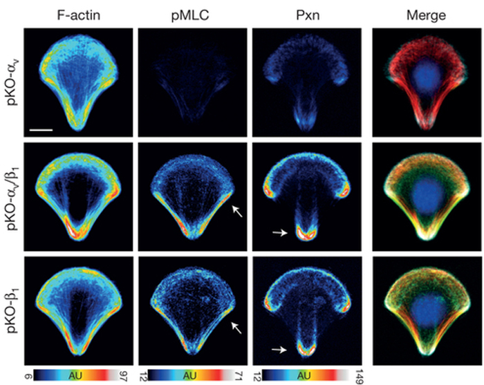
Integrins are transmembrane proteins that attach a cell to another cell or its surrounding extracellular matrix, so their role in regulating cell adhesion in normal cell function, development, and disease is of serious interest to biologists. There are several types of integrins, frequently expressed together at the same time. A recent paper describes a system to investigate how specific integrins function in cell adhesion, and found notable differences. Schiller and colleagues expressed either β1- or αv-class integrins in fibroblasts lacking all other integrins, and found that β1-class integrins are important in small peripheral adhesions, while αv-class integrins are important for large focal adhesions. In the images above, activity of the actin-based motor myosin and presence of the focal adhesion protein paxillin are shown in cells plated on micropatterned shapes. Cells with both β1- and αv-class integrins (middle row) have the highest myosin activity (pMLC) and paxillin signal, while cells with either β1- or αv-integrin alone have lower myosin activity and paxillin signal.
Adapted by permission from Macmillan Publishers Ltd, copyright ©2013
April 5, 2013
Cadherins are transmembrane proteins that form cell-cell adhesion structures called adherens junctions. There are several types of adherens junctions, but they are all composed of clusters of cadherins whose extracellular domains interact with other cells’ cadherins and intracellular domains interact with the cell’s cytoskeleton. Individual cadherin molecules provide negligible adhesive properties, so understanding how cadherin clusters form is an important question. A recent paper delves into the details of this process, and finds that actin filaments are indeed necessary for cadherin cluster stability. Hong and colleagues found that cadherin clusters that were uncoupled from actin were unstable and exhibited random mobility. When the actin-binding domain of a cadherin-actin adaptor protein called α-catenin (domain called αABD) was coupled to these mutant cadherin structures, the adhesive clusters regained stability and deliberate mobility. The images above show clusters of this αABD-cadherin chimera (left, green in merged) associated with actin filaments (middle, red in merged; arrows in inset point to colocalization).
October 23, 2012
The progression of tumor cells to metastatic cancer cells correlates with poor prognoses for cancer patients. The steps that drive cancer cells to spread (metastasis) are not well understood, but may be effective targets for chemotherapies. For example, tumor cells lose adhesion to their underlying extracellular matrix (ECM) prior to spreading to other regions of the body. Understanding this loss of cell-ECM adhesion may guide the development of new therapies. A recent paper describes the systematic analysis of cell-ECM adhesion in tumor cells by using robotically spotted arrays of 768 paired combinations of ECM molecules. Reticker-Flynn and colleagues monitored the adhesion profiles of lung cancer cell lines at different stages of cancer progression on these arrays of ECM molecules, and found ECM-cell interactions that may be successful therapeutic targets. The images above show an array of spotted ECM protein combinations (visible through immuno- and fluorescent-labeling, top), and examples of cells adhered to the ECM spots (bottom images).
Adapted by permission from Macmillan Publishers Ltd, copyright ©2012
June 7, 2012
April 12, 2012
“Hang in there!” says the kitten dangling from a tree branch. Maybe this poster from my junior high-era bedroom should have had a white blood cell instead. While defending the body from infection, white blood cells have to anchor themselves to avoid being swept away from the constant blood flow around them. Today’s image is from a recent paper showing how this happens. Leukocytes, or white blood cells, find their way to sites of infection in the body. Once there, leukocytes are subjected to the force of blood flow around them and must resist detachment from the inflamed tissue. Integrin cell adhesion proteins are important in stabilizing the anchors formed on leukocytes recruited to inflamed tissue. A recent paper shows that rapid actin polymerization at adhesion sites is triggered by the force of blood flow. In addition, Rullo and colleagues show that this actin polymerization is necessary for successful attachment. Image above shows human leukocytes on a surface coated with VCAM-1, a leukocyte adhesion molecule, and exposed to a fluid flow in the direction of the arrow. Arrowhead points to anchor points.
March 12, 2012
If it walks like a duck and quacks like a duck, it must be a duck….or it could be me doing my best duck impression. Scientists don’t like to make assumptions, but instead are driven to painstakingly test the cellular unknowns. Today’s image is from a paper that serves as a good example of how assumptions may not be accurate, but instead lead to a fascinating story that prompts more questions.Desmosomes are cell-cell adhesion structures that help tissues resist mechanical stress by connecting with the cells’ intermediate filament networks. The mechanical strength provided by desmosomes is well documented in tissues such as heart muscle and stratified epithelia (skin, esophagus, etc). The intestine is a simple epithelial tissue that contains desmosomes, and as it is under a lot of mechanical stress from the movement and content of digesting food, it can be easily assumed that the desmosomes in intestinal tissue provide mechanical support. Not so, according to a recent paper by Sumigray and Lechler. The desmosomal protein desmoplakin (DP) is not essential for cell adhesion or tissue integrity in intestinal epithelia, nor is it necessary for proper organization of keratin intermediate filaments. However, DP is important for the structure of microvilli, the actin-rich structures that provide surface area for the absorption of nutrients. As seen in the images above, the microvilli of intestinal cells from DP knockout mice (middle, right) are shorter and misshapen when compared to wild-type cells (left).
February 6, 2012
Watching your child learn to crawl, you realize how much coordination she needs to get up on all fours and move forward, working both sides of her body. You are convinced that your child is totally gifted and brilliant. Well, I have news for you…cells have a lot more to sort out in order to crawl. As you sheepishly compare your child’s brilliance to a cell and admit defeat (except for me, of course…my daughter really IS brilliant), take a moment to look at today’s beautiful images from a paper on cell migration. When a cell is crawling, it first reaches out using membrane protrusions. At the leading edge of these protrusions, the cell will adhere to the underlying matrix. These nascent adhesions serve as anchors to the surface and give the crawling cell traction. Cell-matrix adhesions go through dynamic cycles of formation as nascent adhesions, maturation into focal adhesions, and turnover using a well-studied set of cytoskeletal proteins and regulators, but how these adhesions form and mature is not completely understood. Lawson and colleagues recently published results showing that a protein called FAK (focal adhesion kinase) promotes the recruitment of an adhesion protein called talin to nascent adhesions. Talin binds to integrin, a key adhesion protein, and was previously thought to recruit FAK to nascent adhesions. In the images above, a control cell (left) shows localization of talin (green) to nascent adhesions (red). However, without FAK (right), talin is not recruited to nascent adhesions.
January 26, 2012
There are so many images that are in our collective memory…images that mark historic and significant events. There are the photos of Tiananmen Square, the “Migrant Mother” from the Great Depression, Abbey Road, etc. Well, cell biologists have our own images that stick in our collective memory. One of those more recent images is the “Svitkina image” of actin filaments, which I’ve mentioned before. So, when I saw that the Svitkina lab published a paper recently, I knew I had to share!Cell-cell junctions are crucial for development, tissue structure, and cell-cell communication. One type of cell-cell junction is the adherens junction (AJ), which is a cadherin-based junction that links to the actin cytoskeleton within the cell. Although AJs are well-studied structures, how they assemble is still not completely known. A recent paper looks at the underlying actin filaments in developing AJs. According to Hoelzle and Svitkina, a junction is formed first by neighboring cells’ lamellipodia, sheet-like membrane extensions. Next, the two cells are connected by cadherin on thin bridges that look similar to filopodia, which are finger-like actin projections. Interestingly, these bridges form by actin filament growth from the rear-side of the lamellipodia towards the cell periphery. The images above are transmission electron micrographs of actin filaments in a bridge that connects two different cells (each cell labeled a different color in middle image). Gold beads (yellow, right image) found at the far ends of each cell’s bridge label VASP proteins, which are markers for filopodia.
January 5, 2012
Despite my two-year old daughter’s observation that gummy fruit snacks are great adhesive tools, the tissues in our body require something a bit more sophisticated to stick together. Different types of tissue need different specialized adhesion structures. For example, desmosomes function in heart and skin tissue, which are under a lot of mechanical stress. Today’s image is from a paper describing how some desmosome proteins get to the adhesion site. Desmosomes are highly-ordered structures at the plasma membrane that adhere cells to one another, and play a crucial role in maintaining tissue integrity both during and after development. The adhesion properties of desmosomes are due to the presence of two different cadherin proteins, called Dsg and Dsc. A recent paper describes how these two cadherins are trafficked to desmosome adhesion sites. According to Nekrasova and colleagues, Dsg and Dsc are transported to desmosomes by two different kinesins, which are motors that walk along microtubules. Dsg is transported by kinesin-1, while Dsc is transported by kinesin-2. That each desmosome cadherin has its own transport pathway suggests that the assembly and function of desmosomes, and in turn adhesion, can be tailored throughout development and tissue remodeling. In the sequence of images above, Dsg (red, arrow) is migrating along microtubules (blue) towards the cell periphery.
September 22, 2011
I love it when worlds collide. I love the movies where a country boy falls for a city girl. Or a robot develops a friendship with a wookie. Hilarity typically ensues in the movies, but fantastic new ideas and questions result from the discovery of biological processes colliding. So, when I came across a recent paper that revealed new results on the relationship between endocytosis and adhesion, I was all over it. Cell-cell adhesion is constantly adjusted throughout development, wound healing, and cancer metastasis. E-cadherin is the major adhesion molecule that functions in epithelial cell adhesion and polarity, and is linked to the actin skeleton (via α-catenin) and p120. The level of E-cadherin at the cell surface influences the adhesive strength between two cells, and this strength can be adjusted by internalization (endocytosis) of E-cadherin away from the cell surface. A recent paper discusses results showing how internalization of E-cadherin is regulated by Numb, a protein that interacts with endocytosis adaptor proteins and is important throughout development. Sato and colleagues found that Numb interacts directly with p120, and showed that impairment of Numb prevents E-cadherin internalization. The images above show cysts of epithelial cells. In control cysts (top rows), E-cadherin and p120 (red) were found at the basolateral cell-cell junctions. In cysts with reduced levels of Numb (bottom rows), both E-cadherin and p120 localized to the apical membrane region (blue) too.
August 18, 2011
I bet you think you’re pretty good at wearing the many proverbial hats in your life. I bet you can align a laser, walk your dog, change a diaper, and play in your awesome band of cell biologists. Well, integrins will put your hat-wearing to shame! Integrins are very important proteins (VIPs!) that play huge roles in adhesion, signaling, polarity, cell migration, cell division, and differentiation. Today’s image is from a paper describing new data on integrin trafficking.
Integrins are membrane proteins that interact with the environment outside of the cell to regulate cell adhesion and signaling. As part of the cell’s plasma membrane, integrins are constantly being brought into the cell and recycled back to the cell surface. Understanding this process is important—the way that integrins are recycled back to the cell’s surface (or not) can dramatically affect a cell’s ability to move, adhere to other cells, divide, and invade (in the case of cells in a tumor). A recent paper by Mai and colleagues describes a protein called RASA1 in regulating integrin recycling back to the membrane. RASA1 binds to integrin on a site where another protein called Rab21 also binds. So, these two proteins compete—Rab21 bound to integrin prevents its recycling back to the cell surface, while RASA1 binding allows integrin to traffic back to the surface. In the images above, when levels of RASA1 were reduced (bottom), cells were able to migrate more efficiently to close a “wound” scratched across a layer of cells, as compared to control cells (top). Images on the left show the wound shortly after it was created, while images on the right are four hours later.
July 7, 2011
Totally tubular! If Bill and Ted had an excellent adventure in the human body, you can be certain that they’d learn about the most excellent tube structures throughout the body. From the veins that carry our blood to the branching tubules in our lungs, tubes are very important structures. A recent paper looks at the role of adhesion proteins during tubule formation.During development, dramatic rearrangements of epithelial sheets results in the formation of branched tubules, as seen in kidney, lung, and mammary gland tissue. As one might expect, these rearrangements require coordination of several cellular events such as cell division, migration, polarization, and adhesion. A recent paper describes the role of two adhesion proteins, E-cadherin and cadherin-6, in tubule formation. Jia and colleagues found that cadherin-6 is important in inhibiting tubule formation, while E-cadherin is important in the formation of a tubule’s lumen (its inside cavity). Images above show the use of cell cysts as a model for epithelial tubule and lumen formation, with fluorescent tags showing a lateral marker (blue) and lumen-facing apical markers (green and red). Samples of control cysts, cysts without cadherin-6, E-cadherin, or both are shown (moving left to right). Although the mutant cysts appear abnormal, polarization was not disrupted in cysts without either cadherin (although multiple lumens are visible in cysts lacking E-cadherin). The polarization of cysts lacking both cadherins, however, was completely disrupted.
February 14, 2011
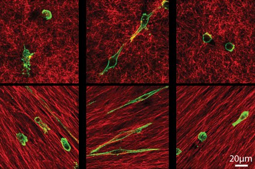
Cancer cells are smart little guys, unfortunately. Many cancer therapies target a specific type of cancer cell migration, but many cancer cells are able to switch to a different mode of migration and evade the attack. Today’s image is from a paper looking at these two modes of migration by using 3D cultures of malignant breast cancer cells.Malignant cancer cells are able to spread beyond the initial tumor, and their migration can occur from using either of two types of cancer cell migration—mesenchymal and amoeboid. Mesenchymal migration is characterized by elongated cancer cells that can undergo proteolysis of and adhesion to the extracellular matrix on which it is migrating. Cancer cells undergoing amoeboid migration are rounded and squeeze through extracellular spaces without proteolysis of or adhesion to the matrix. A recent paper looks at the distinct roles of two different adhesion adaptor proteins – paxillin and Hic-5 – in these two modes of migration. Images above are of breast cancer cells (green) grown in two different types of 3D cultures (top and bottom). Compared with the mixed morphologies of control breast cancer cells (left images), cells without paxillin (middle images) had elongated, mesenchymal morphologies. Breast cancer cells without Hic-5 (right images) had more rounded, amoeboid morphologies.
December 9, 2010
Some of the most striking and informative images aren’t of cells or organisms, but are computer-generated representations of what is going on in cells or organisms. These computer-generated images come from the use of two-photon microscopy, a powerful technique that allows for imaging of tissue that’s buried deep in a living organism. Zebrafish is a freshwater fish that serves as a great model organism to cell and developmental biologists. During development, cells undergo dramatic reorganization during formation of the central nervous system, a process called neurulation. A recent paper describes the interaction between two proteins, called Protocadherin-19 and N-cadherin, and how these two proteins regulate cell movements during neurulation. These proteins together regulate cell-cell adhesion at a time when cells converge together to form a midline seam in the neural plate, a key feature of brain development. Images above are cell trajectories taken from time-lapse series of zebrafish embryos undergoing neurulation. The trajectories of cells in a normal embryo (top left) show a convergence of cells to the midline and a general movement of cells in one direction. Cells showed compromised movement in embryos with low levels of either protocadherin-19 (top, right), N-cadherin (bottom, left), or both (bottom, right).
BONUS!! Cool movies of two-photon image sequences can be found here.
November 29, 2010
Basic research is fundamental if we want to make strides in understanding disease. Please (politely) shout this from the mountaintops and make sure your lawmakers and funding agencies understand this. Today’s image is from a paper that investigates a key player in cell scattering, which is an event very similar to cancer metastasis. Cell scattering is a term used to describe cell-cell dissociation and migration and occurs in liver development, organ regeneration, and metastasis. Cell scattering is induced by hepatocyte growth factor/scatter factor (HGF/SF1), and a recent paper describes similar cell scattering after a block to the protein α5β1 integrin, which is a receptor mediating the attachment of the cell to the surrounding extracellular matrix. In addition, blocking the function of this integrin triggers changes in expression of other proteins that mediate adhesion and migration. Image shows control (top) or integrin-blocked (bottom) liver progenitor cells. After a functional block to integrin function, cells are scattered and have decreased levels of E-cadherin (green), which is important in cell-cell adhesion of epithelial cells. Nuclei are in blue.
September 23, 2010

Retinal detachment occurs when the retinal pigment epithelium (RPE) loses its adhesion to the underlying photoreceptors, and is a sight-threatening condition. A recent paper has looked at the role of a protein called CLIC4 in this adhesion, and found decreased retinal adhesion in CLIC4-suppressed RPE cells. Images are of normal (top) and CLIC4-suppressed (bottom) retinal sections, showing the disorganization of the photoreceptors in the outer nuclear layer (ONL) of retinas with CLIC4-suppressed RPE cells.
Reference: Jen-Zen Chuang, Szu-Yi Chou, and Ching-Hwa Sung. Authors’ Molecular Biology of the Cell paper can be found here.
September 6, 2010
Strong cell-cell adhesion is crucial for tissue organization during development. A complex of three proteins—cadherin, α-catenin, and β-catenin—play an important role in adhesion by organizing and regulating the actin cytoskeleton. A recent paper demonstrates how α-catenin functions within this complex and with actin in the developing worm embryo, and shows that this complex is regulated differently from the mechanism in mammals. Image is a C. elegans embryo with a mutant form of α-catenin (blue and green) and actin (yellow and magenta). Both are localized at cell junctions in normal embryos, but in this mutant there is reduced α-catenin and gaps of actin localization at cell-cell junctions.Reference: Image is by Stephanie L. Maiden, and is the cover image cover for the August issue of PNAS, which can be found here. Accompanying paper is by Adam V. Kwiatkowski, Stephanie L. Maiden, Sabine Pokutta, Hee-Jung Choi, Jacqueline M. Benjamin, Allison M. Lynch, W. James Nelson, William I. Weis, and Jeff Hardin, and can be found here.