Showing posts with label clathrin. Show all posts

January 10, 2013

The idea of screening for something valuable is something we’ve all done. When looking for a dog several years ago, I screened through PetFinder to find the exact dog I wanted to bring home based on size, age, scruffiness, etc. Despite the fact that this dog just farted at my feet, my PetFinder screen brought me to my best friend. For a biologist, screening can lead to exciting discoveries about what genes are important in a specific process (minus the gas, I think).

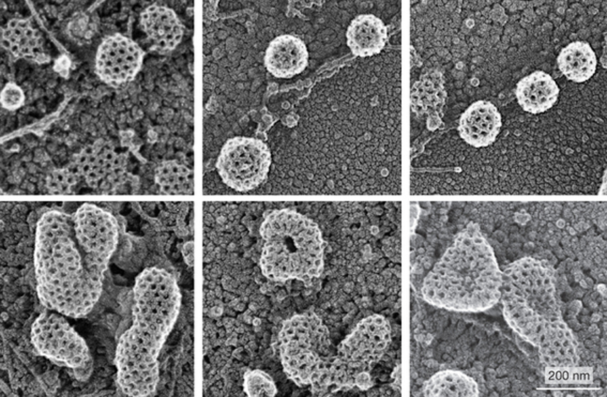
During endocytosis, a cell takes in material from its outside environment. Clathrin-mediated endocytosis involves the inward budding of vesicles, and depends on a lattice, or “coat”, of clathrin molecules that help shape the rounded vesicle. A recent paper describes the results of a screen to find regulators of clathrin-coated vesicle formation at the plasma membrane, one of the earliest steps in clathrin-mediated endocytosis. Kozik and colleagues screened the entire human genome for regulators, and found 92 genes that affect this process. One of these genes encodes the protein V-ATPase, which is found in many membranes and can pump protons across a membrane to regulate pH. In the images above, V-ATPase-inhibited cells (bottom) formed enlarged clathrin-coated structures, compared to the small and uniform clathrin-coated vesicles in control cells (top). Kozik and colleagues found that V-ATPase inhibition blocked the recycling of cholesterol back to the plasma membrane, where it has been suggested that cholesterol aids in membrane bending.

 BONUS!! Electron microscopy image of a heart-shaped vesicle, acquired as part of the screen to find clathrin regulators.

ResearchBlogging.orgKozik, P., Hodson, N., Sahlender, D., Simecek, N., Soromani, C., Wu, J., Collinson, L., & Robinson, M. (2012). A human genome-wide screen for regulators of clathrin-coated vesicle formation reveals an unexpected role for the V-ATPase Nature Cell Biology, 15 (1), 50-60 DOI: 10.1038/ncb2652  
Adapted by permission from Macmillan Publishers Ltd, copyright ©2013

June 21, 2012

There’s a song on the radio that you just love right now. You’re not sure what it is about the song that you love…the catchy chorus, the singer’s silky voice, the booming bass line, the touch of pan flute, whatever. If you’re musically-inclined, you start teasing the song apart in your head until you finally realize what it is (always, it’s the pan flute!). Knowing how to tease a complex problem apart is a key skill for any scientist, and today’s image is from a paper that pares down clathrin-coated pits to their bare minimum.

Clathrin coats are assembled cages of the scaffolding protein clathrin. These scaffolds deform a planar membrane into a curved membrane that is able to bud off during the uptake of material into the cell, a process called endocytosis. It was previously thought that clathrin-associated proteins helped to induce curvature in the membrane, but recently a group finds otherwise, in a study that describes the minimum requirements for clathrin-coated bud formation. In this paper, Dannhauser and Ungewickell monitored bud and vesicle formation using a cell-free system composed of brain lipids that artificially form vesicles called liposomes. They found that clathrin alone is sufficient to generate buds in a lipid membrane. In the images above, buds and vesicles were formed when liposomes were in the presence of clathrin, but lacking any other proteins able to induce membrane curvature. Many vesicles were, in fact, still attached to the liposomes via narrow membrane necks (arrows). 

ResearchBlogging.orgPhilip N. Dannhauser, & Ernst J. Ungewickell (2012). Reconstitution of clathrin-coated bud and vesicle formation with minimal components Nature Cell Biology, 14 (6), 634-639 : 10.1038/ncb2478
Adapted by permission from Macmillan Publishers Ltd, copyright ©2012

May 31, 2012

I love it when things really throw me for a loop. Like when I heard that Kim Kardashian earned a perfect SAT score and was accepted into Harvard. Okay, that’s not really true…but just imagine our collective “whaaaa?!” In science there are always things that flip your lid, and today’s image is from a paper that does just that. Clathrin-coated pit closure just got more interesting.**

Clathrin-mediated endocytosis is a process during which material is brought into the cell through pits coated with a clathrin lattice, which provides structure to the developing vesicle. Electron microscopy images of clathrin-coated pits show great detail about their structure, but can only provide snapshots of what occurs inside of the cell. A recent paper describes clathrin-coated pit closure using a technique that combines the imaging of structural detail seen in electron microscopy (in this case, called scanning ion conductance microscopy) with live confocal microscopy used to track fluorescently-tagged proteins. During conventional clathrin pit closure, the pit is closed and cleaved from flat membrane sheets. Shevchuk and colleagues, however, found that 70% of pits close using an alternative mechanism. In this mechanism, a membrane protrusion grew from one side of the clathrin pit and covered the pit to close it, as seen in the images above.

**Halvsies if this tagline prompts next summer’s blockbuster movie. 

ResearchBlogging.orgShevchuk, A., Novak, P., Taylor, M., Diakonov, I., Ziyadeh-Isleem, A., Bitoun, M., Guicheney, P., Lab, M., Gorelik, J., Merrifield, C., Klenerman, D., & Korchev, Y. (2012). An alternative mechanism of clathrin-coated pit closure revealed by ion conductance microscopy originally published in the Journal of Cell Biology, 197 (4), 499-508 DOI: 10.1083/jcb.201109130

June 23, 2011

Life is a balance of giving and taking, and this starts with our cells. I’ve written about cells taking in material (endocytosis) plenty of times, but it’s time to talk about cells secreting material out of the cell. Check out today’s stunning image of salivary gland cells in the fruit fly larva.

All cells undergo some base level of secretion, but there are many cell types with specialized “regulated” secretion. For example, our endocrine cells secrete the hormones that regulate our bodies and throw teenagers into crazed states. Cells with regulated secretion store high concentrations of certain proteins in dense organelles called secretory granules, until there is a signal that triggers the release of these proteins. A recent paper asks how secretory granules are formed, and finds that two vesicle coat proteins, called AP-1 and clathrin, are required. Burgess and colleagues looked at secretory granules in larval fruit fly salivary glands, and found that AP1 and clathrin are localized at newly synthesized secretory proteins, Golgi structures (where the proteins are sorted), and maturing secretory granules. Images show salivary gland cells with AP1 (red) colocalizing with Golgi structures (green).

ResearchBlogging.orgBurgess, J., Jauregui, M., Tan, J., Rollins, J., Lallet, S., Leventis, P., Boulianne, G., Chang, H., Le Borgne, R., Kramer, H., & Brill, J. (2011). AP-1 and clathrin are essential for secretory granule biogenesis in Drosophila Molecular Biology of the Cell DOI: 10.1091/mbc.E11-01-0054

June 7, 2010


Endocytosis, the uptake of material into a cell, often involves the structural protein clathrin. Clathrin forms a curved lattice on the plasma membrane that buds inward and eventually pinches off, carrying the material in a clathrin-coated vesicle. Last year, a group carefully measured the assembly and dynamics of clathrin structures and found that there are two distinct clathrin structures that are regulated differently, and interact with different cellular structures. Clathrin coated pits are the rapidly forming and sharply curved canonical structures of clathrin-mediated endocytosis, while clathrin coated plaques are longer-lived and less sharply curved structures. Electron microscopy image above shows both clathrin coated pits (top row) and plaques (bottom) in HeLa cells.

Reference: Saveez Saffarian, Emanuele Cocucci, Tomas Kirchhausen. Authors’ PLoS Biology paper can be found here.