I’m willing to bet that most scientists were pretty destructive as kids. Not intentionally destructive, though…I bet we all liked taking things apart to see what each part of a toy did. Maybe your Teddy Ruxpin eventually sounded like a demonic doll after your experiments, or you finally pulled apart your Etch-A-Sketch to uncover the magic. Either way, it was early training for what scientists do every day to understand cells better. Today’s image is from a paper that takes this route to understand cell adhesion better.
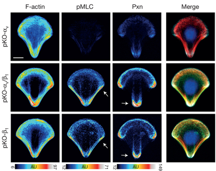
Integrins are transmembrane proteins that attach a cell to another cell or its surrounding extracellular matrix, so their role in regulating cell adhesion in normal cell function, development, and disease is of serious interest to biologists. There are several types of integrins, frequently expressed together at the same time. A recent paper describes a system to investigate how specific integrins function in cell adhesion, and found notable differences. Schiller and colleagues expressed either β1- or αv-class integrins in fibroblasts lacking all other integrins, and found that β1-class integrins are important in small peripheral adhesions, while αv-class integrins are important for large focal adhesions. In the images above, activity of the actin-based motor myosin and presence of the focal adhesion protein paxillin are shown in cells plated on micropatterned shapes. Cells with both β1- and αv-class integrins (middle row) have the highest myosin activity (pMLC) and paxillin signal, while cells with either β1- or αv-integrin alone have lower myosin activity and paxillin signal.
 Schiller, H., Hermann, M., Polleux, J., Vignaud, T., Zanivan, S., Friedel, C., Sun, Z., Raducanu, A., Gottschalk, K., Théry, M., Mann, M., & Fässler, R. (2013). β1- and αv-class integrins cooperate to regulate myosin II during rigidity sensing of fibronectin-based microenvironments Nature Cell Biology, 15 (6), 625-636 DOI: 10.1038/ncb2747
Adapted by permission from Macmillan Publishers Ltd, copyright ©2013
Schiller, H., Hermann, M., Polleux, J., Vignaud, T., Zanivan, S., Friedel, C., Sun, Z., Raducanu, A., Gottschalk, K., Théry, M., Mann, M., & Fässler, R. (2013). β1- and αv-class integrins cooperate to regulate myosin II during rigidity sensing of fibronectin-based microenvironments Nature Cell Biology, 15 (6), 625-636 DOI: 10.1038/ncb2747
Adapted by permission from Macmillan Publishers Ltd, copyright ©2013
 I bet you think you’re pretty good at wearing the many proverbial hats in your life. I bet you can align a laser, walk your dog, change a diaper, and play in your awesome band of cell biologists. Well, integrins will put your hat-wearing to shame! Integrins are very important proteins (VIPs!) that play huge roles in adhesion, signaling, polarity, cell migration, cell division, and differentiation. Today’s image is from a paper describing new data on integrin trafficking.
Integrins are membrane proteins that interact with the environment outside of the cell to regulate cell adhesion and signaling. As part of the cell’s plasma membrane, integrins are constantly being brought into the cell and recycled back to the cell surface. Understanding this process is important—the way that integrins are recycled back to the cell’s surface (or not) can dramatically affect a cell’s ability to move, adhere to other cells, divide, and invade (in the case of cells in a tumor). A recent paper by Mai and colleagues describes a protein called RASA1 in regulating integrin recycling back to the membrane. RASA1 binds to integrin on a site where another protein called Rab21 also binds. So, these two proteins compete—Rab21 bound to integrin prevents its recycling back to the cell surface, while RASA1 binding allows integrin to traffic back to the surface. In the images above, when levels of RASA1 were reduced (bottom), cells were able to migrate more efficiently to close a “wound” scratched across a layer of cells, as compared to control cells (top). Images on the left show the wound shortly after it was created, while images on the right are four hours later.
Mai, A., Veltel, S., Pellinen, T., Padzik, A., Coffey, E., Marjomaki, V., & Ivaska, J. (2011). Competitive binding of Rab21 and p120RasGAP to integrins regulates receptor traffic and migration originally published in The Journal of Cell Biology, 194 (2), 291-306 DOI: 10.1083/jcb.201012126