A personal note

This will be my last HighMag post for a while, as I await the August arrival of my second daughter.  While I love looking at beautiful images of cells and reading exciting new papers, I am currently obsessed with insane nesting projects…as if the baby will be interested in our how organized our garage is.  After Baby arrives and my sleep deprivation eases up a bit (ha!), I look forward to posting again on HighMag.

To make sure you don’t miss future HighMag posts, be sure to “Like” the HighMag Facebook page.  All of the cool kids are doing it, so you should too.

If you need to contact me for anything, feel free to email me at highmagblog@gmail.com.  

Until I’m back in the HighMag saddle, check out these HighMag Greatest Hits I've compiled for you.  The following are links to posts that were either your favorite images and papers (lots of traffic!) or my own personal favorites.  Enjoy!

2013

2012

2011

2010

July 17, 2013

We don’t need to reinvent the wheel (even if someone tried to in 2001...click here).  We use the wheel for so many things ranging from transport to energy.  Cells have proven clever at co-opting machinery for multiple processes, as the paper from today’s image describes.  This recent paper shows the use of specific machinery in both cytokinesis and neuronal migration.

When neurons migrate, there is a leading process in the front of the cell body and a trailing process.  The leading process contains actin filaments that enable the cell body of the neuron to move forward.  A recent paper describes how the microtubule-based motor kinesin-6 plays an important role in neuronal migration.  Kinesin-6 is best known for its role in cytokinesis, the physical division of a cell after mitosis.  Falnikar and colleagues found that kinesin-6 concentrates in the same region as actin filaments in the leading process of a migrating neuron.  Without kinesin-6, neurons lose their bipolar leading-trailing process morphology, concentrate actin filaments in more than one process, and either remain stationary or continually change the direction of migration.  In addition, Falnikar and colleagues found that kinesin-6 signals through the GTPase activating protein MgcRacGAP to regulate the actin cytoskeleton, as it does during cytokinesis.  In the images above, control neurons (top time-lapse series) moved in a single direction, while neurons depleted of kinesin-6 bottom) frequently changed directions.

BONUS!!  Check out a movie of a wandering, migrating kinesin-6-depleted neuron below.

ResearchBlogging.orgAditi Falnikar, Shubha Tole, Mei Liu, Judy S. Liu, & Peter W. Baas (2013). Polarity in Migrating Neurons Is Related to a Mechanism Analogous to Cytokinesis Current Biology, 23 (13), 1215-1220 DOI: 10.1016/j.cub.2013.05.027 Copyright ©2013 Elsevier Ltd. All rights reserved.

July 10, 2013

The grace of a migrating cell is as deceiving as a pair of Spanx on an English Bulldog… there is a lot going underneath.  Today’s image is from a paper showing the importance of the protein vinculin at the leading edge of a migrating cell.

Cell migration is driven by actin filament polymerization that pushes the leading edge of the cell forward, as well as F-actin retrograde flow.  Focal adhesions (FAs) adhere the crawling cell to the underlying extracellular matrix (ECM), and are assembled and disassembled near the leading edge of the cell.  Proteins of these FAs are believed to make up a “molecular clutch” that engages the retrograde F-actin flow, and a recent paper identifies the protein vinculin, an actin-binding protein, as a molecular clutch.  Thievessen and colleagues investigated the effects of vinculin gene disruption in migrating fibroblasts, and found that vinculin is important in regulating F-actin organization and FA dynamics.  Specifically, vinculin generates the ECM traction forces necessary for migration, and promotes FA formation and turnover.  In the images above, a normal fibroblast (top) and a fibroblast lacking vinculin (bottom) show F-actin (green) and the lamellipodial protein cortactin (purple).  Normal crawling fibroblasts have a sharply defined band of cortactin colocalized with F-actin at the leading edge, while vinculin mutants have a wider, less defined region of cortactin at the leading edge, suggesting the importance of vinculin in leading edge organization.

BONUS!  Check out some cool movies from this paper here.

ResearchBlogging.orgThievessen I, Thompson PM, Berlemont S, Plevock KM, Plotnikov SV, Zemljic-Harpf A, Ross RS, Davidson MW, Danuser G, Campbell SL, & Waterman CM (2013). Vinculin-actin interaction couples actin retrograde flow to focal adhesions, but is dispensable for focal adhesion growth. originally published in the Journal of Cell Biology, 202 (1), 163-77 PMID: 23836933

July 2, 2013

 
Timing is everything….from the fluke encounter in a romantic comedy, to your rush to make the bus/train/plane this morning, to the development of an organism. Today’s image is from a paper describing the temporal patterning involved in the development of the fruit fly optic lobe.

In the fruit fly optic lobe, the medulla processes visual information using 40,000 neurons of over 70 different cell types. The medulla develops from a crescent-shaped tissue from which the neuronal progenitors divide, requiring several different transcription factors. A recent paper describes the sequential patterning of five transcription factors as the medulla neuroblasts age. Li and colleagues found that this temporal patterning of transcription factors is necessary for the diversity of cell types found in the medulla. The images of the developing medulla above show the sequential expression of these five transcription factors—Homothorax (Hth), Eyeless (Ey), Sloppy paired 1 and 2 (Slp), Dichaete (D), and Tailless (Tll)—in five consecutive stripes. Hth is found in the youngest neuroblasts. Ey, Slp, and D are required for turning on the next transcription factor in the cascade. Slp and D are also required for turning off the preceding transcription factor.

ResearchBlogging.orgLi X, Erclik T, Bertet C, Chen Z, Voutev R, Venkatesh S, Morante J, Celik A, & Desplan C (2013). Temporal patterning of Drosophila medulla neuroblasts controls neural fates. Nature, 498 (7455), 456-62 PMID: 23783517 
Adapted by permission from Macmillan Publishers Ltd, copyright ©2013

June 25, 2013

Maybe you’ve been cruising in your minivan blasting “The Wheels on the Bus” too loudly for your preschooler, or maybe you’re preparing to hear the witching hour’s cries of a newborn due soon (this blog is way too autobiographical).  Either way, our ears endure a lot of stress from loud sounds throughout our lives.  The images above are from a paper describing a mechanism for handling this stress.

Our inner ear hair cells detect sound when stereocilia projections are deflected by sound vibrations.  These actin-based projections are connected to each other by tiny extracellular tip links, which are tugged when the stereocilia bend.  The tugging of these tip links is believed to drive the opening of transduction channels that result in the electrical signals relayed to the auditory nerve.  Although mammalian hair cells do not regenerate over the course of a lifetime, the tip links do.  A recent paper investigates the mechanism behind tip link regeneration.  Indzhykulian and colleagues developed an electron microscopy technique to visualize the tip links on hair cell stereocilia and immuno-gold labeled proteins of tip links, and found a two-step mechanism for tip link regeneration.  This mechanism begins with shorter tip links composed of just protocadherin 15, and follows with mature tip links containing protocadherin 15 and cadherin 23.  In the images above, stereocilia tip links were regenerated after nearly complete tip disruption by the chemical BAPTA.  Insets show higher magnified views of the tip links, which appear regenerated by 24-48 hours, compared with control tip links (top) and BAPTA-treated cells (second row).

ResearchBlogging.orgIndzhykulian AA, Stepanyan R, Nelina A, Spinelli KJ, Ahmed ZM, Belyantseva IA, Friedman TB, Barr-Gillespie PG, & Frolenkov GI (2013). Molecular remodeling of tip links underlies mechanosensory regeneration in auditory hair cells. PLoS biology, 11 (6) PMID: 23776407

June 21, 2013

Good things come in small packages.  Maybe I’m referring to the burst of antioxidants jammed into tiny blueberries.  Maybe I’m referring to my tiny three year-old who yells, “Come oooon, THAT was funny!” when I don’t laugh loudly enough at her jokes.  Or maybe I’m referring to C. elegans.  These worms are tiny, but pack a serious punch of significant biology that helps us learn about important cellular processes.  Today’s image is from a paper that serves as an excellent example of this.

During cell invasion, a cell is able to breach and cross over the basement membrane that underlies a sheet of epithelial cells.  Cell invasion occurs throughout development and in the spread of cancer, yet biologists studying cell invasion have been challenged by the difficulty of visualizing the event.  A recent paper describes the development of live-cell imaging methods for studying cell invasion, using the worm’s anchor cell.  The anchor cell in the developing worm’s uterus breaches the basement membrane in order to link uterine and vulval tissues, and its transmigration is precisely timed.  Hagedorn and colleagues followed the interactions between the invading anchor cell and the basement membrane, and found very dynamic actin-based invadopodia that first breach the basement membrane.  These protrusions then stabilize to expand the breach and cross into the vulval tissue.  Anchor cell invasion depends on the netrin receptor UNC-40 (DCC) at the interface between the anchor cell and basement membrane.  In the time-lapse images above, the invading protrusion (cyan in top, grayscale in bottom) can be seen breaching the basement membrane (purple) and invading the vulval tissue underneath.

ResearchBlogging.orgHagedorn, E., Ziel, J., Morrissey, M., Linden, L., Wang, Z., Chi, Q., Johnson, S., & Sherwood, D. (2013). The netrin receptor DCC focuses invadopodia-driven basement membrane transmigration in vivo originally published in the Journal of Cell Biology, 201 (6), 903-913 DOI: 10.1083/jcb.201301091

June 18, 2013

The study of how cells move in development is not just about development.  Understanding cell migration can also help researchers understand how tumors spread and invade other tissues.  So, the next time you see someone roll their eyes at your fruit fly egg chambers (or worm vulva, or culture dishes), take pity at their ignorance and explain to them how they should thank you instead.

The movement of cells during development drives the shape changes and organization of an embryo.  In the fruit fly ovary, a small cluster of border cells migrates across a region of the egg chamber in order to reach the oocyte.  This collective migration of these border cells depends on polarization of the actin cytoskeleton.  A recent paper describes the role of the Hippo signaling pathway in driving the polarization of actin to the outer rim of the migrating border cell cluster.  Lucas and colleagues found that upstream Hippo pathway components localize to the contacts between border cells within the cluster in order to link polarity signaling with actin cytoskeleton organization.  In the images above, the actin cytoskeleton (red) can be seen at the outer rim of the migrating cluster of border cells (arrows) as it moves across the egg chamber towards the oocyte (top to bottom, chronologically).  Higher magnification views of the cluster are on the right.

 ResearchBlogging.orgLucas, E., Khanal, I., Gaspar, P., Fletcher, G., Polesello, C., Tapon, N., & Thompson, B. (2013). The Hippo pathway polarizes the actin cytoskeleton during collective migration of Drosophila border cells originally published in the Journal of Cell Biology, 201 (6), 875-885 DOI: 10.1083/jcb.201210073

June 11, 2013

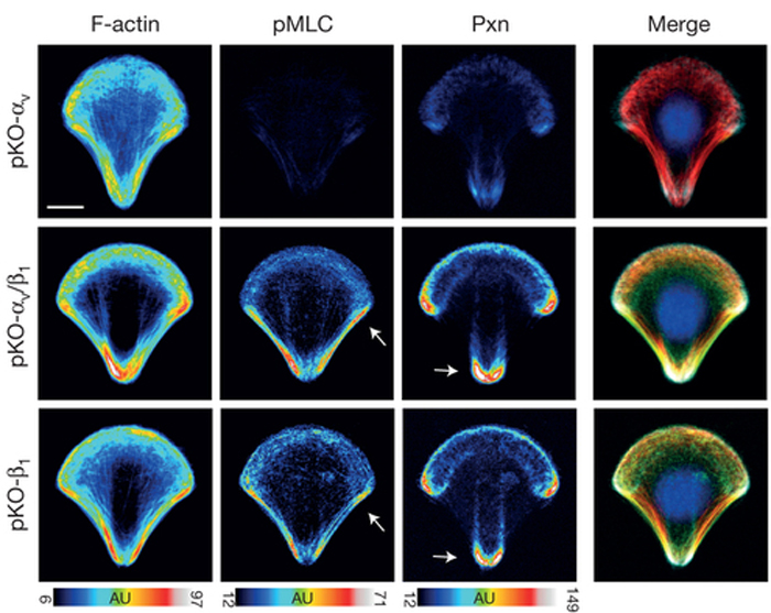
 I’m willing to bet that most scientists were pretty destructive as kids.  Not intentionally destructive, though…I bet we all liked taking things apart to see what each part of a toy did.  Maybe your Teddy Ruxpin eventually sounded like a demonic doll after your experiments, or you finally pulled apart your Etch-A-Sketch to uncover the magic.  Either way, it was early training for what scientists do every day to understand cells better.  Today’s image is from a paper that takes this route to understand cell adhesion better.

Integrins are transmembrane proteins that attach a cell to another cell or its surrounding extracellular matrix, so their role in regulating cell adhesion in normal cell function, development, and disease is of serious interest to biologists.  There are several types of integrins, frequently expressed together at the same time.  A recent paper describes a system to investigate how specific integrins function in cell adhesion, and found notable differences.  Schiller and colleagues expressed either β1- or αv-class integrins in fibroblasts lacking all other integrins, and found that β1-class integrins are important in small peripheral adhesions, while αv-class integrins are important for large focal adhesions.  In the images above, activity of the actin-based motor myosin and presence of the focal adhesion protein paxillin are shown in cells plated on micropatterned shapes.  Cells with both β1- and αv-class integrins (middle row) have the highest myosin activity (pMLC) and paxillin signal, while cells with either β1- or αv-integrin alone have lower myosin activity and paxillin signal.  

ResearchBlogging.orgSchiller, H., Hermann, M., Polleux, J., Vignaud, T., Zanivan, S., Friedel, C., Sun, Z., Raducanu, A., Gottschalk, K., Théry, M., Mann, M., & Fässler, R. (2013). β1- and αv-class integrins cooperate to regulate myosin II during rigidity sensing of fibronectin-based microenvironments Nature Cell Biology, 15 (6), 625-636 DOI: 10.1038/ncb2747
Adapted by permission from Macmillan Publishers Ltd, copyright ©2013 

June 4, 2013

HighMag is back from an early summer vacation at the beach, and ready to get our microscopic groove on.  Happy Summer, everyone!


Our world is the same size it’s always been, but so many advances in technology have made the world seem a lot smaller.  We can call our brother across the country and video chat with our sister on the other side of the world, all while searching the internet for the lyrics to Snow’s “Informer” (side note…knowing the lyrics won’t help you understand the song AT ALL).  Similarly, a neuron’s axons make the nervous system seem a lot smaller too, with their amazing ability to grow very long.  Today’s image is from a paper showing how microtubule sliding is involved.

Neurons can transmit signals to far away neurons, thanks to the ability of their axons to grow insanely long.  Both actin filaments and microtubules participate in axon growth, with microtubules pushing the growing axon out and actin filaments working directly at the tip of the growing axon, called the growth cone.  A recent paper shows the role of microtubule sliding in the initial growth of neurites in fruit fly neurons.  Lu and colleagues found that the microtubule motor kinesin-1 drives the sliding of microtubules past one other in order to push out the neuron’s growing projection.  This mechanism does not require actin filaments, and is suppressed during maturation of neurons.  In the images above, microtubules (green) can be seen pushing against the tip of a growing neurite in a young neuron (membrane of neuron in red and bottom right series).  The whole neuron is on the left, and the neurite in the boxed region is shown in the time-lapse images on the right.

ResearchBlogging.orgLu, W., Fox, P., Lakonishok, M., Davidson, M., & Gelfand, V. (2013). Initial Neurite Outgrowth in Drosophila Neurons Is Driven by Kinesin-Powered Microtubule Sliding Current Biology, 23 (11), 1018-1023 DOI: 10.1016/j.cub.2013.04.050 
Copyright ©2013 Elsevier Ltd. All rights reserved.

May 24, 2013

There isn’t a cell biologist out there who doesn’t fantasize about reaching her hands into a cell and physically manipulating whatever protein or structure that she obsesses over.  While we can’t do that with our own hands, optical tweezers can…and the information we learn is invaluable.  Today’s image is from a paper that uses optical tweezers to measure the forces within a mitotic spindle.

The mechanics within a mitotic spindle are complicated, and cannot be fully understood until all of the forces that exist within it are determined.  A recent paper measures the forces of chromosome and spindle pole movements using optical tweezers to trap either kinetochores or spindle poles.  Optical tweezers use refracted light to trap small objects within a cell, and are able to stop the movement of the object as the laser power of the trap approaches (or exceeds) the cell’s own force on the object.  With this technology, Ferraro-Gideon and colleagues measured the forces in mitotic spindles in several samples—a flatworm, a crane fly, and a mammalian spindle.  The force used to stop chromosome movement was closer to the theoretical values of chromosome force, much less than the force values calculated by experiments performed by Bruce Nicklas in the 1980s using needles to manipulate chromosome movement.  In the DIC (left) and fluorescence images (right) above, a trap (square) is applied to kinetochores in mammalian cells.  The trap did not damage the microtubules of the spindle (right image).

ResearchBlogging.orgFerraro-Gideon, J., Sheykhani, R., Zhu, Q., Duquette, M., Berns, M., & Forer, A. (2013). Measurements of forces produced by the mitotic spindle using optical tweezers Molecular Biology of the Cell, 24 (9), 1375-1386 DOI: 10.1091/mbc.E12-12-0901

Check out an extended report on these experiments, and others from this lab:
Sheykhani, R., Baker, N., Gomez-Godinez, V., Liaw, L., Shah, J., Berns, M., & Forer, A. (2013). The role of actin and myosin in PtK2 spindle length changes induced by laser microbeam irradiations across the spindle Cytoskeleton, 70 (5), 241-259 DOI: 10.1002/cm.21104

May 21, 2013

“LET THERE BE LIGHT!” said the microscopist.  Light plays a crucial role in microscopy and cell biology, and a recent paper describes the use of light to understand protein secretion.

Light is used in microscopy in countless ways—to illuminate a sample, excite a fluorophore, and signal the localization or dynamics of a protein.  Light can also be used to manipulate cellular events through the use of “caged” compounds that become active after illumination by certain wavelengths of light.  This technology gives biologists the ability to spatially and temporally control cellular events in order to understand them better.  Recent advances in this technology use illumination of plant photoreceptors to control protein-protein interactions, but some cellular processes such as protein secretion have been difficult to manipulate.  A recent paper describes the use of the plant photoreceptor UVR8 in the first light-triggered protein secretion system developed.  Chen and colleagues have shown that the recently described UVR8 can conditionally sequester proteins bound for secretion in the ER, and then upon illumination with UV light releases these proteins to the plasma membrane.  In the images above, a neuron before (left) and after (right) UV illumination with this UVR8 system shows the movement of proteins known to be secreted from the soma and dendritic processes (arrowheads), where the ER is distributed, and into the Golgi (arrow), a necessary step in protein secretion.

ResearchBlogging.orgChen, D., Gibson, E., & Kennedy, M. (2013). A light-triggered protein secretion system originally published in the Journal of Cell Biology, 201 (4), 631-640 DOI: 10.1083/jcb.201210119

May 13, 2013

Molecular motors are some of the raddest things in a cell.  They can walk along cytoskeletal elements such as microtubules and actin filaments, and the list of cellular events that they participate in is a long, long list.  Today’s image is from a paper showing a beautiful pattern of nonmuscle myosin II in epithelial cells.

Epithelial cells assemble junctions to adhere to one another, and the actin motor nonmuscle myosin II (NMII) is a major component of these epithelial apical junctions.  NMII helps the epithelial sheet respond to morphogenesis and changes in tissue homeostasis, and a recent paper describes how the network of NMII motors does so.  Ebrahim and colleagues have found that NMII in the apical junctional complex of epithelial cells assembles into precise muscle-like sarcomere units that form a belt around each cell.  The sarcomeres of neighboring cells are aligned, in turn assembling into a contractile network that can result in changes in cell shape.  In the images above, NMII (green) is seen in repeated sarcomere units around each cell (actin is in red).  NMII puncta are paired together in neighboring cells.  Arrows (middle) point to the junctions between three cells, seen at higher magnification on the right.

ResearchBlogging.orgEbrahim, S., Fujita, T., Millis, B., Kozin, E., Ma, X., Kawamoto, S., Baird, M., Davidson, M., Yonemura, S., Hisa, Y., Conti, M., Adelstein, R., Sakaguchi, H., & Kachar, B. (2013). NMII Forms a Contractile Transcellular Sarcomeric Network to Regulate Apical Cell Junctions and Tissue Geometry Current Biology, 23 (8), 731-736 DOI: 10.1016/j.cub.2013.03.039 
Copyright ©2013 Elsevier Ltd. All rights reserved.

May 9, 2013

When we think of wounds, we don’t typically think of them as part of normal, healthy function.  Micro-wounds, however, form when white blood cells have to cross the barrier in our blood vessels to get to an injury or infection.  These micro-wounds happen all the time, and our cells heal these wounds efficiently and elegantly.

One of the most important barriers in our body is that created by the vascular endothelium.  Vascular endothelial cells line all of our blood vessels—from the largest vessels to the smallest capillaries—and function in fluid filtration, hormone trafficking, and recruitment and trafficking of blood and stem cells.  The movement of cells, for example white blood cells, across the vascular endothelium and out of circulation creates “micro-wounds” that can compromise the integrity of the tissue.  A recent paper describes how these micro-wounds are healed, based on a model in which the vascular endothelium senses a loss of tension upon micro-wounding and triggers its own repair.  Martinelli and colleagues tracked micro-wounds that were created by either transmigrating white blood cells or by mechanical disruption by a probe, and found that ventral lamellipodia are generated by endothelial cells to close the micro-wounds.  These lamellipodia are enriched in Rac1 effector proteins, and require reactive oxygen species (ROS) and Arp2/3 for efficient wound closing.  Images above show probe-induced wounding of an endothelial cell, followed by wound healing.  The wound initially expanded to 20um across (80 seconds), with multiple nodes of ventral lamellipodia (blue arrowheads) and ventral F-actin waves forming around the wound and closing it.

ResearchBlogging.orgMartinelli, R., Kamei, M., Sage, P., Massol, R., Varghese, L., Sciuto, T., Toporsian, M., Dvorak, A., Kirchhausen, T., Springer, T., & Carman, C. (2013). Release of cellular tension signals self-restorative ventral lamellipodia to heal barrier micro-wounds originally published in the Journal of Cell Biology, 201 (3), 449-465 DOI: 10.1083/jcb.201209077

May 6, 2013

Our bodies have multiple fronts for battling viruses, and it’s impressive that any of those suckers manage to invade our bodies at all.  When virus particles do make their way into a cell, it’s important for biologists to understand their pathway through a cell in order to create drug therapies and vaccines.  Today’s image is from a paper describing the use of high resolution imaging to understand this process.

The polarized cells that line our digestive and respiratory tracts form a tight barrier that protects our bodies from viruses.  Understanding how viruses are able to breach these polarized epithelial cells is important in guiding the development of therapeutics and vaccines, yet previous research has focused mainly on in vitro studies of virus entry into nonpolarized cells.  Microscopy advances have recently allowed the high-resolution imaging of virus entry in polarized epithelial cells.  Boulant and colleagues used live-cell spinning-disk confocal microscopy to follow the uptake of single mammalian reovirus (MRV) virions and infectious subvirion particles (ISVPs) in polarized Madin–Darby canine kidney cells.  Both virus particles were internalized by clathrin-mediated endocytosis at the apical surface.  MRV virions reached early and late endosomes, while ISVPs escaped the endocytic pathway prior to reaching early endosomes.  In the images above, the tight-junction protein ZO-1 (red, left) shows the typical belt pattern surrounding the polarized cells (side views are also shown in the top and side strips).  The MRV cell surface receptor JAM-A (red, right) is localized near tight junctions and on the apical side of the cells.

ResearchBlogging.orgBoulant, S., Stanifer, M., Kural, C., Cureton, D., Massol, R., Nibert, M., & Kirchhausen, T. (2013). Similar uptake but different trafficking and escape routes of reovirus virions and infectious subvirion particles imaged in polarized Madin-Darby canine kidney cells Molecular Biology of the Cell, 24 (8), 1196-1207 DOI: 10.1091/mbc.E12-12-0852

May 2, 2013

If cells had their own soundtracks, I think any flagella-wielding cells would take home the prize.  Maybe the soundtrack begins on a high note with Devo’s “Whip It!”, continues on to the more crass Clarence Carter, plateaus with some headbanging death metal, and finally pays homage to Flock of Seagulls simply due to their flagellar waveform hair.  Today’s image is from a paper describing an outer-inner dynein link in flagella.

Flagella are whip-like organelles protruding from cells, and function to move fluid past the cell to generate motility.  In flagella, nine doublets of microtubules are bundled around a central microtubule pair (the 9+2 structure).  Outer dynein arms (ODAs) and inner dynein arms (IDAs) drive movement of the microtubule doublets past each other, generating the flagellar beating motion.  The ODA and IDA play distinct roles in flagellar function, but a recent paper finds a link between them.  Oda and colleagues found that intermediate chain 2 (IC2) of ODAs functions as part of the outer-inner dynein linker.  IC2 is a hub between ODAs and IDAs to regulate flagellar beating, based on the beating motion of IC2 mutants in the green algae Chlamydomonas.  The images above show the waveforms and image sequences of swimming Chlamydomonas cells.  The bending of flagella in wild-type cells (top row) is different from that of ic2 mutant cells (bottom row).  The principle bend at the flagellar tip in the forward stroke persists in the mutant (arrowhead); this altered bending results in slower swimming for the mutants.

ResearchBlogging.orgOda, T., Yagi, T., Yanagisawa, H., & Kikkawa, M. (2013). Identification of the Outer-Inner Dynein Linker as a Hub Controller for Axonemal Dynein Activities Current Biology, 23 (8), 656-664 DOI: 10.1016/j.cub.2013.03.028
Copyright ©2013 Elsevier Ltd. All rights reserved.

April 29, 2013

There are many, many things in cell biology that can serve as models for fine art, but fewer are more stunning to me than a Purkinje neuron.  Purkinje neurons are some of the largest neurons in the brain, where they participate in motor control from the cerebellum.  Today’s image is from a paper describing what happens when a protein called rictor is depleted from Purkinje neurons.

The two multi-protein complexes mTORC1 and mTORC2 share sensitivity to inhibition by the immunosuppressive drug rapamycin, yet likely have distinct roles in cell function and development.  Each complex is composed of a distinct set of subunits—mTORC1 depends on the protein raptor, while mTORC2 depends on the protein rictor.  A recent paper describes an important role for mTORC2 in neuron size, morphology, and function.  Thomanetz and colleagues found that two different mouse lines lacking the mTORC2 protein rictor had smaller neurons with disrupted function, likely mediated through regulation of PKC (protein kinase C) isoforms.   mTORC1 activity was unaffected in these mutants.  When rictor was depleted from the entire central nervous system, motor function of the mice was affected.  When rictor was depleted from only Purkinje neurons, the cell type with the highest rictor expression, neurons were smaller and their morphology was abnormal.  In the images above, control Purkinje neurons (top) had only one primary dendrite compared with neurons lacking rictor (bottom), which had multiple primary dendrites (notated in different colors, right).  

Thomanetz, V., Angliker, N., Cloetta, D., Lustenberger, R., Schweighauser, M., Oliveri, F., Suzuki, N., & Ruegg, M. (2013). Ablation of the mTORC2 component rictor in brain or Purkinje cells affects size and neuron morphology originally published in the Journal of Cell Biology, 201 (2), 293-308 DOI: 10.1083/jcb.201205030ResearchBlogging.org

April 26, 2013

It’s Worm Week here at HighMag Blog.  Worms are amazing little creatures, and the species C. elegans is an invaluable model system for studying cell and developmental biology.  Their genome is sequenced, their development is precise and well-documented, and their bodies and embryos are translucent (making them photogenic under a microscope).  Today’s image is from the same lab that brought Tuesday’s image…worm gonads rock!

Blurb and image from Christian R. Eckmann:
The image is an immuno-stained part of an extruded C. elegans hermaphrodite gonad; germ cell nuclei (magenta) and the apical membrane (green). The germ stem cells reside at the closed end of this tube like tissue. In wild type, the germ stem cells exit the mitotic zone, entering meiosis further away from the closed tip and start differentiating into sperm or oocytes. 

The image posted earlier this week is from the Gracida and Eckmann paper that identifies a nuclear receptor that protects germ stem cell integrity, and in turn fertility, after dietary perturbations.

Gracida, X. & Eckmann, C. (2013). Fertility and Germline Stem Cell Maintenance under Different Diets Requires nhr-114/HNF4 in C. elegans Current Biology, 23 (7), 607-613 DOI: 10.1016/j.cub.2013.02.034 


April 23, 2013

I’m thankful that my body knows how to handle days when I feed it wonderful things, like a banana and a giant bowl of strawberries, then follow it up with a few gut-busting mini-doughnuts.  Although worms and other organisms don’t have access to doughnuts like I do, their bodies still have protections in place to handle changes in their diet.  Today’s image is from a paper describing how the germline is protected from a changing diet.

Organisms consume a variety of food options, yet their bodies know how to regulate these changes to maintain homeostasis, all the way down to the cellular level.  A recent paper shows how an organism’s germline stem cells (GSCs), the source of eggs and/or sperm, are protected from food intake.  Gracida and Eckmann found that the nuclear receptor NHR-114 protects GSCs from dietary perturbations in worms, possibly through a detoxifying response to certain food intake.  Without NHR-114, worms on certain bacterial diets become sterile due to germ cell division defects during development.  The dietary sensitivity is based on intake of the amino acid tryptophan.  In the images above, gonads of worms fed a certain bacterial diet are stained to see individual germ cells (cell cortex staining in green in merged; DNA is purple).  Compared to wild-type worms (left), worms depleted of NHR-114 (right) have germ cell defects, notably cells with multiple nuclei (arrowhead).

ResearchBlogging.orgGracida, X., & Eckmann, C. (2013). Fertility and Germline Stem Cell Maintenance under Different Diets Requires nhr-114/HNF4 in C. elegans Current Biology, 23 (7), 607-613 DOI: 10.1016/j.cub.2013.02.034 
Copyright ©2013 Elsevier Ltd. All rights reserved. 

April 16, 2013

I’m as type-A as a person can get, with my organized desk, to-do lists, and inability to roll with it (I sound dreadful, right?).  One thing that will never change is the calm I feel when I read the word “systematic” in a paper’s title or abstract.  Anything done systematically soothes me like a heartbeat soothes a newborn.  Today’s image is from a paper that uses a—you guessed it—systematic approach to understanding the roles of dynein and its many regulators in mitosis.

Dynein is a large microtubule motor complex that is important in countless cellular processes, most notably mitosis.  Like most proteins, dynein relies on numerous adaptor proteins and the dynactin complex to help localize the motor and/or activate it.  A recent paper uses siRNA screening to systematically test the roles of dynein subunits, adaptor proteins, and dynactin subunits to build a more complete picture of the roles of each protein in mitosis.  Raaijmakers and colleagues show that while some regulators are required for activation but not localization of dynein, others are required mainly for dynein localization.  Dynactin, for example, is not necessary for spindle organization, but rather serves as a dynein recruitment factor at the nuclear envelope and kinetochores.  In the images above, the mitotic spindle in a control cell is very focused at spindle poles (second row, green in merged).  When the dynein heavy chain subunit is depleted from cells (all other columns), spindles show a range of spindle pole focusing defects, including spindles lacking attachment to their poles.  Spindle microtubules are top row, red in merged; chromosomes are third row, blue in merged.

ResearchBlogging.orgRaaijmakers, J., Tanenbaum, M., & Medema, R. (2013). Systematic dissection of dynein regulators in mitosis originally published in the Journal of Cell Biology, 201 (2), 201-215 DOI: 10.1083/jcb.201208098

April 12, 2013

I think I speak for many when I say that dinosaurs were the first objects of our life-long science obsessions.  Their size, history, and ferocious good looks fascinate even the youngest preschoolers.  Although my obsession turned to microscopic things, some folks remained true to their love of dinosaurs and history.  Today’s image is a treat, and a great example of how the basic questions in developmental biology know no timeline.

The study of dinosaur embryos at the cellular levels helps scientists understand the growth patterns of dinosaurs.  Despite this, little is known about dinosaur embryos, as they are rare and typically still inside of their eggshells.  A recently discovered dinosaur embryo bone bed in China is the oldest dinosaur embryo find in fossil record, from the Lower Jurassic about 190-200 million years ago.  These embryos represent several different nests, and are at different developmental stages.   In addition, these embryos are likely that of a Lufengosaurus, from the sauropodomorph clade of dinosaurs known for long necks and gigantism.  Because of the variation of age of these found embryos, Reisz and colleagues were able to track development through several different stages, including very early embryonic stages.  In the images above, thin sections from three different femur (thigh) bone samples (of 24 discovered total) show changes in the bone tissue formation as the embryos age from youngest (left) or oldest (right).  Two different regions of the femora are shown for each sample (top and bottom).

BONUS!  More thin sections are shown below.

ResearchBlogging.orgReisz, R., Huang, T., Roberts, E., Peng, S., Sullivan, C., Stein, K., LeBlanc, A., Shieh, D., Chang, R., Chiang, C., Yang, C., & Zhong, S. (2013). Embryology of Early Jurassic dinosaur from China with evidence of preserved organic remains Nature, 496 (7444), 210-214 DOI: 10.1038/nature11978 
Adapted by permission from Macmillan Publishers Ltd, copyright ©2013 

April 9, 2013

Our genome is chock full of so many things that aren’t even genes.  In fact, only about 2% of the human genome actually encodes protein sequences...mind blown, right?!  There are many different kinds of elements and domains within our genome that regulate gene expression through their roles in chromosome architecture and organization.  Today’s image is from a paper that describes the dynamics of one type of domain—the lamina associated domain.

The nuclear lamina is a protein layer that coats the inside nuclear membrane, and serves to anchor chromosomes.  Regions in the genome called lamina associated domains (LADs) specifically associate with the nuclear lamina.  LADs cover about 35-40% of the genome, suggesting that they may affect chromosome position and architecture.  A recent paper tracks LAD-nuclear lamina interactions throughout the cell cycle in single cells.  Kind and colleagues show that about 30% of LADs are positioned at the nuclear periphery.  LADs are stochastically positioned after mitosis, meaning that their position is not directly inherited.  In addition, these contacts are linked with gene expression and histone modifications.  In the images above, the nuclear lamina (blue) and mitotic spindle (red) are shown throughout the different stages of mitosis.  LADs (green) are rounded and at the nuclear periphery during prophase, and then become banded along chromosomes once the nuclear envelope breaks down (prometaphase and metaphase).  During cytokinesis, LADs are seen within the nucleus, but not yet positioned at the periphery.

ResearchBlogging.orgKind, J., Pagie, L., Ortabozkoyun, H., Boyle, S., de Vries, S., Janssen, H., Amendola, M., Nolen, L., Bickmore, W., & van Steensel, B. (2013). Single-Cell Dynamics of Genome-Nuclear Lamina Interactions Cell, 153 (1), 178-192 DOI: 10.1016/j.cell.2013.02.028
Copyright ©2013 Elsevier Ltd. All rights reserved.

April 5, 2013

Cell adhesion is sticky business.  See what I did there?!  Comedy. Gold.  Seriously, though, cell adhesion is complicated, with many types of cell adhesion structures that form at specific regions of the cell at specific times.  As important as it is to understand cell adhesion and its role in development, cancer, and normal cell function, we are all thankful for papers like the one that today’s image comes from.

Cadherins are transmembrane proteins that form cell-cell adhesion structures called adherens junctions.  There are several types of adherens junctions, but they are all composed of clusters of cadherins whose extracellular domains interact with other cells’ cadherins and intracellular domains interact with the cell’s cytoskeleton.  Individual cadherin molecules provide negligible adhesive properties, so understanding how cadherin clusters form is an important question.   A recent paper delves into the details of this process, and finds that actin filaments are indeed necessary for cadherin cluster stability.  Hong and colleagues found that cadherin clusters that were uncoupled from actin were unstable and exhibited random mobility.  When the actin-binding domain of a cadherin-actin adaptor protein called α-catenin (domain called αABD) was coupled to these mutant cadherin structures, the adhesive clusters regained stability and deliberate mobility.  The images above show clusters of this αABD-cadherin chimera (left, green in merged) associated with actin filaments (middle, red in merged; arrows in inset point to colocalization).

ResearchBlogging.orgHong, S., Troyanovsky, R., & Troyanovsky, S. (2013). Binding to F-actin guides cadherin cluster assembly, stability, and movement originally published in the Journal of Cell Biology, 201 (1), 131-143 DOI: 10.1083/jcb.201211054

April 2, 2013

As I write this, I have dirt underneath my fingernails and I love it.  Spring is here, and I have begun playing in the dirt and cheering for my budding vegetable garden seedlings.  I love the food plants provide us, but they’re also fascinating models for understanding cell biology and developmental biology.  Today’s image is from a paper identifying a player in the development of stomata, which are important plant organs.

Stomata are pore organs on leaves that regulate gas and water vapor exchange in plants.  They are made of pairs of guard cells that regulate the size of the stomata openings to let air in and oxygen out.  A recent paper describes the identification of a protein that regulates the maturing and functioning of stomatal guard cells.  Negi and colleagues identified SCAP1, a transcription factor, that when mutated results in irregularly-shaped guard cells.  These mutants also lack the ability to control stomatal opening and closing.  SCAP1 regulates the transcription of known guard cell development genes.  The images above show a wild-type plant (top) with normal developing stomata at all stages (mature stomata is right-most image).  In a scap1 mutant (bottom), however, later stages of stomata development are defective and result in stomata with a floppy or irregular appearance.

ResearchBlogging.orgNegi, J., Moriwaki, K., Konishi, M., Yokoyama, R., Nakano, T., Kusumi, K., Hashimoto-Sugimoto, M., Schroeder, J., Nishitani, K., Yanagisawa, S., & Iba, K. (2013). A Dof Transcription Factor, SCAP1, Is Essential for the Development of Functional Stomata in Arabidopsis Current Biology, 23 (6), 479-484 DOI: 10.1016/j.cub.2013.02.001
Copyright ©2013 Elsevier Ltd. All rights reserved.